

A 5-year-old boy presented with lesions on his left cheek and temporal area that had appeared a few days earlier. Each lesion started as an erythematous papule and evolved into a coin-sized ulcerated plaque. The plaques featured brown, central adherent crusts and yellow-brown, dried marginal exudates with surrounding erythema (Figure 1).
FIGURE 1
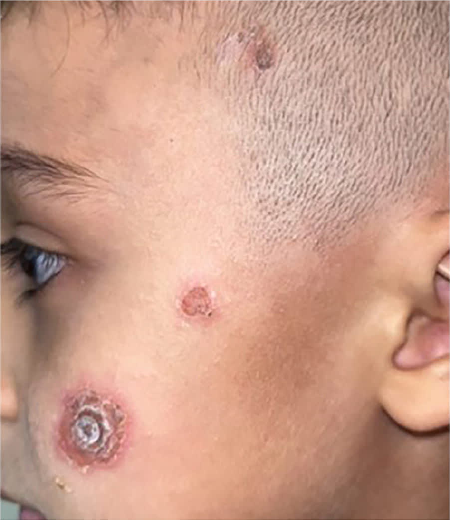
The patient did not have a history of insect or arthropod bites, and he did not report pain, pruritus, or associated fever or chills. Fusidic acid (a topical antibacterial) was applied to the lesions, but this did not lead to improvement. The patient was then prescribed cephalexin (50 mg/kg per day). One week later, the lesions had improved significantly.
QUESTION
Based on the patient's history and physical examination, which one of the following is the most likely diagnosis?
- A. Ecthyma.
- B. Ecthyma gangrenosum.
- C. Impetigo.
- D. Pyoderma gangrenosum.
DISCUSSION
The answer is A: ecthyma. This ulcerative bacterial skin infection is caused by group A streptococci and is often associated with secondary staphylococcal infection. Ecthyma typically begins superficially and extends into the dermis. It can be referred to as a deeper form of impetigo. However, impetigo affects only the stratum corneum, whereas ecthyma extends into the dermis and results in scarring.1
Ecthyma lesions manifest as small, fluid-filled vesicles or pustules that ulcerate and leave a thick gray-yellow crust. If the crust is removed, a superficial ulcer is revealed with a depressed, raw base and raised edges. The ulcers heal within a few weeks but often leave scarring. Ecthyma most commonly occurs on the lower extremities and in patients at the extremes of age. Immuno-suppression, poor hygiene, overcrowding, malnutrition, high humidity, and preexisting trauma to the tissue are predisposing factors. If large areas are involved or the lesions do not respond to topical antibiotics, systemic antibiotics with good gram-positive coverage should be prescribed.2
Ecthyma gangrenosum is a pseudomonal infection manifesting as a papule with a hemorrhagic crust. Lesions are usually larger than 3 cm in diameter. It results from the hematogenous spread of Pseudomonas aeruginosa. The condition usually occurs in patients who are immunosuppressed or extremely ill. When ecthyma gangrenosum is suspected, biopsy with deep-tissue Gram stain and culture can be obtained for diagnosis.3
Impetigo is a common skin infection caused by staphylococcal or streptococcal bacteria. It occurs most often in children. The rash typically begins as red papules that become vesicles and develop into crusted lesions. The crusting is typically honey colored, not red or brown, and erythema usually is not present. The paranasal skin is a common site for the rash, although it can occur anywhere.4
Pyoderma gangrenosum is an inflammatory, noninfectious, ulcerative neutrophilic skin disease of uncertain etiology. It is commonly misdiagnosed as an aggressive skin infection. Typically, a single pustule evolves into a large ulcer with a necrotic and undermined margin. Pyoderma gangrenosum can have an acute or chronic course. It often results in extensive scarring, which can be keloidal or dyspigmented, especially in patients with darker skin types. It can affect patients of any age but is most common in middle-aged adults.5
Read the full article
Get immediate access, anytime, anywhere.
Choose a single article, issue, or full-access subscription.
Earn up to 14 CME credits per issue.
