

A 68-year-old man presented with black discoloration of his left great toe and toenail that had been present for more than 1 year. The toe was not painful. Treatment with antifungal spray did not lead to improvement. The patient did not have a history of injury or trauma to the toe.
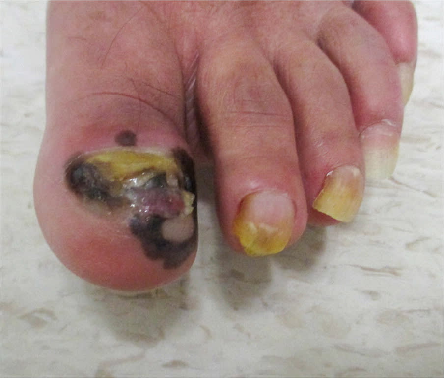
Physical examination revealed a large, irregularly pigmented black and brown plaque of the subungual skin from the left great toe to the proximal and medial nail folds (Figure 1). The toenail was almost completely destroyed, and a large pink nodule with purulent discharge was present in the nail bed. Pinprick sensation was decreased on the plantar surface of the first and second toes.
FIGURE 1
QUESTION
Based on the patient's history and physical examination, which one of the following is the most likely diagnosis?
- A. Diabetic foot ulcer.
- B. Longitudinal melanonychia striata.
- C. Onychomycosis.
- D. Pyogenic granuloma.
- E. Subungual melanoma.
DISCUSSION
The answer is E: subungual melanoma. Sixty-five percent of cases present as a longitudinal band of nail discoloration with proximal widening and irregular side borders. The nail plate may also thicken or split.1 Patients may present with diffuse melanonychia, nail dystrophy, nail bed lesions or ulceration, subungual masses, osseous involvement, and lack of gross pigmentation. Hutchinson sign (brown-black pigmentation of subungual skin extending to proximal and lateral nail folds) is associated with subungual melanoma, with sensitivity of 42% and specificity of 96%.2,3 However, it may also occur with subungual hematoma, fungal infection, and drug-induced dyschromia.4,5
The extended ABCDEF mnemonic summarizes common features of subungual melanoma6:
- Age: in patients 50 to 70 years
- Brown or black, breadth more than 3 mm, irregular borders
- Change in nail band or morphology despite adequate treatment
- Digit: the great toe is most commonly involved
- Extension of pigment onto proximal and lateral nail fold
- Family history of dysplastic nevus or melanoma
Subungual melanoma is a rare subtype of acral-lentiginous melanoma (0.7% to 3% of all melanomas). Acral-lentiginous melanomas may be more common among African American, Asian, and Hispanic people; however, studies did not address how race and ethnicity were defined, which limits this data.7
Among cutaneous melanomas, acral-lentiginous melanomas are associated with the poorest prognosis, partly due to its aggressive nature and delays in diagnosis and treatment.8 Survival rates vary and depend on nodal involvement, ulceration, and pathologic features such as tumor thickness (ie, Breslow thickness) and mitotic rate. Five-year, recurrence-free survival for locally invasive melanoma ranges from 53% to 80%.9
Diabetic foot ulcers may present as painless chronic ulceration at weight-bearing areas (eg, heel, plantar metatarsal head) and are associated with loss in distal pulses and muscle atrophy. They typically occur in patients with long-standing diabetes and peripheral neuropathy. Macular hyperpigmentation with irregular borders is not a typical finding in diabetic foot disease.10
Longitudinal melanonychia striata is a benign condition that presents as a longitudinal nail band with even pigmentation and width. More than 30% of subungual melanomas begin as longitudinal melanonychia; therefore, patients should be counseled to watch closely for changes.11
Onychomycosis is a fungal infection that typically presents as brittle, thickened, and discolored nails with chalky subungual debris. Although it can present as nail discoloration and destruction in its chronic stages, further workup is needed if the condition progresses despite appropriate antifungal treatment.12
Read the full article
Get immediate access, anytime, anywhere.
Choose a single article, issue, or full-access subscription.
Earn up to 14 CME credits per issue.
