

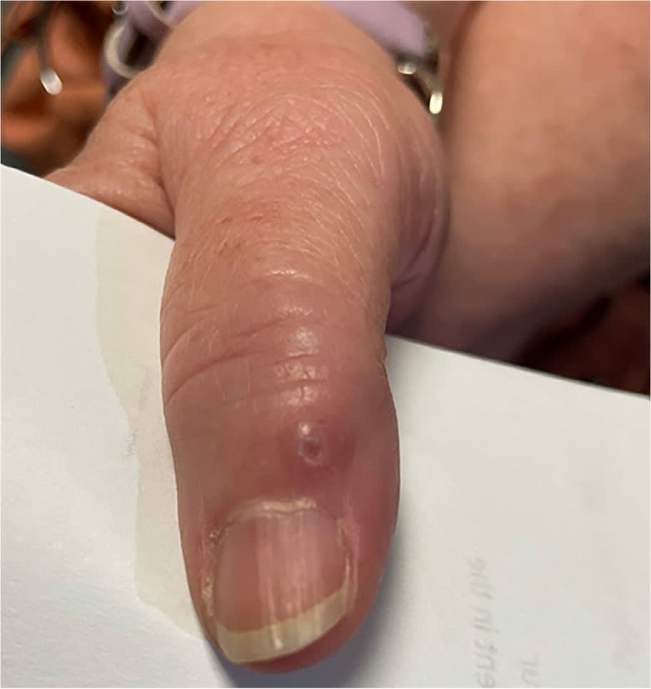
A 61-year-old woman presented with a painful cystic lump on the dorsum of her right thumb. The lump had been present for about 1 year and had slowly increased in size. In the week prior, it became painful and firm. The patient had no other symptoms or lesions. She did not work in a garden, have pets, or experience any trauma to the finger. The lesion had not been treated.
Examination revealed a 3- to 4-mm, raised lesion. It was mildly erythematous with an overlying scab (Figure 1).
FIGURE 1
QUESTION
Based on the patient's history and physical examination, which one of the following is the most likely diagnosis?
- A. Cutaneous sporotrichosis.
- B. Myxoid cyst.
- C. Paronychia.
- D. Verruca vulgaris.
DISCUSSION
The answer is B: myxoid cyst, also known as a myxoid pseudocyst or digital mucous cyst. These benign growths typically present as a single nodule that is translucent or the same color as the surrounding skin. Lesions can also be small, shiny, papular, and fluid-filled. They typically occur on the dorsal aspect of the finger, between the distal interphalangeal joint and the proximal nail fold.1 When the cyst develops close to the proximal nail fold, pressure on the underlying nail matrix can create a groove that runs lengthwise along the nail plate.
Myxoid cysts develop because of mucoid degeneration of connective tissue or the leakage of fluid from an osteoarthritic joint. Radiography of the affected hand can help assess osteoarthritis.2 Treatment of myxoid cysts includes compression, cryotherapy, steroid injection, or surgical excision.3
Cutaneous sporotrichosis, also known as rose gardener's disease, is a fungal infection caused by Sporothrix schenckii. It is associated with gardening or other outdoor activities that expose individuals to contaminated soil or plant matter.4 The infection typically presents as small, painless, pink nodules that may become painful ulcerations. Without treatment, these lesions can spread along lymphatic channels, causing more extensive skin involvement and disseminated cutaneous sporotrichosis.5
Paronychia is a common infection of the skin around the nail. It is caused by infiltration of skin bacteria following disruption of the protective barrier of the nail fold (eg, from nail grooming, nail-biting, thumb-sucking, or habitual hangnail picking).6 Paronychia presents with pain, tenderness, warmth, erythema, and edema in the proximal and lateral nail folds. It may progress to an abscess.7
Verruca vulgaris, also known as the common wart, is a benign cutaneous lesion caused by human papillomavirus infection of keratinocytes. These lesions can manifest on the hands and soles of the feet as small growths that are the same color as the surrounding skin. The growths can be painful when pressure is applied. Verruca vulgaris is spread by contact and can be recognized by its grainy texture.8
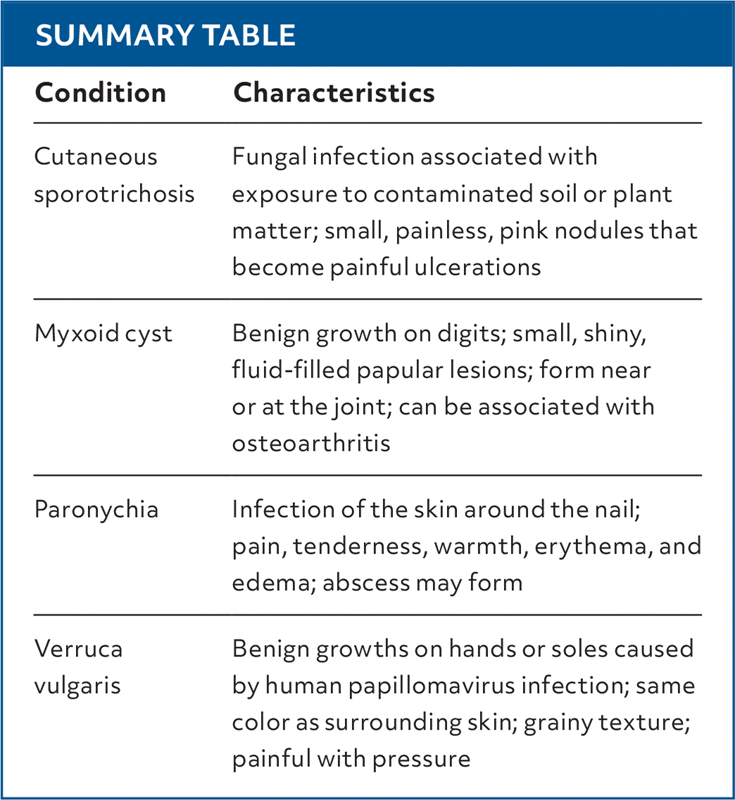
SUMMARY TABLE
| Condition | Characteristics |
|---|---|
| Cutaneous sporotrichosis | Fungal infection associated with exposure to contaminated soil or plant matter; small, painless, pink nodules that become painful ulcerations |
| Myxoid cyst | Benign growth on digits; small, shiny, fluid-filled papular lesions; form near or at the joint; can be associated with osteoarthritis |
| Paronychia | Infection of the skin around the nail; pain, tenderness, warmth, erythema, and edema; abscess may form |
| Verruca vulgaris | Benign growths on hands or soles caused by human papillomavirus infection; same color as surrounding skin; grainy texture; painful with pressure |
Read the full article
Get immediate access, anytime, anywhere.
Choose a single article, issue, or full-access subscription.
Earn up to 14 CME credits per issue.
