

A 64-year-old man presented with a solitary nodule on his chin that had gradually enlarged over several months. The lesion was firm and nontender and did not feature discharge. The patient did not recall any trauma to the area. He did not have other symptoms or a personal or family history of similar skin conditions or autoimmune diseases.
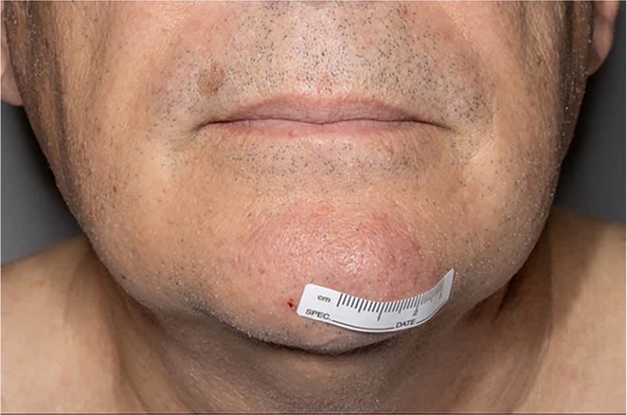
Physical examination revealed a firm, mobile, nonulcerated subcutaneous mass just left of the midpoint on his chin (Figure 1). It was 4 cm in diameter. No other lesions were noted.
FIGURE 1
QUESTION
Based on the patient’s history and physical examination, which one of the following is the most likely diagnosis?
- A. Basal cell carcinoma.
- B. Cutaneous large B-cell lymphoma.
- C. Dermatofibroma.
- D. Epidermal inclusion cyst.
- E. Keratoacanthoma.
DISCUSSION
The answer is B: cutaneous large B-cell lymphoma, an extranodal B-cell non-Hodgkin lymphoma that primarily involves the skin without evidence of extracutaneous disease at the time of diagnosis.1 The etiology is not fully understood; however, it is believed to stem from the clonal proliferation of B cells, genetic abnormalities, chronic inflammation, and immune dysregulation. The condition typically manifests as pruritic, pink to violaceous nodules, plaques, or papules on the scalp, forehead, neck, or trunk. The nodular form progresses slowly with a favorable prognosis, but it has a high rate of recurrence. The diffuse form of cutaneous large B-cell lymphoma has a poor prognosis.2
The patient’s fine-needle aspiration cytology showed atypical lymphocytes. Biopsy demonstrated a 0.6 × 0.4 × 0.1 cm excision of red-brown, rubbery, soft tissue with evidence of a deep, diffuse, and dense infiltrate of B cells with coexpression of the germinal center marker BCL6 and large centrocytic cytology. CD10 and BCL2 were absent. Hematoxylin-eosin staining revealed large lymphocytes with irregular nuclear contours, no nucleoli, and minimal cytoplasm. There was no evidence of granulomas, inclusions, or large noncleaved lymphoid cell components. Positron emission and computed tomography were performed to assess the extent of disease involvement and to evaluate for lymph node enlargement and systemic spread.
First-line treatment for large B-cell lymphoma includes excision or radiotherapy. Depending on the extent of involvement, interferon alfa or intralesional or intravenous anti-CD20 antibodies (eg, rituximab) may also be considered. Chemotherapy is recommended only for extracutaneous spread.2
Basal cell carcinoma is the most common neoplasm of skin and is characterized by pearly papules or nodules with central ulceration. It is common in older patients. Histology shows peripheral palisaded basaloid cells.3
Dermatofibromas are benign fibrous histiocytomas of the skin, usually less than 1 cm in diameter with a characteristic “dimple sign.” The lesions do not cause other symptoms and are related to local trauma. Histology displays proliferation of spindle-shaped fibrous cells mixed with histiocytoid cells in a storiform pattern.4
Epidermal inclusion cysts manifest as benign lesions that are nontender, soft, and mobile. They grow slowly and are more common in adulthood. A keratinized cystic epithelial lining and loose connective tissue capsule are evident on histology.5
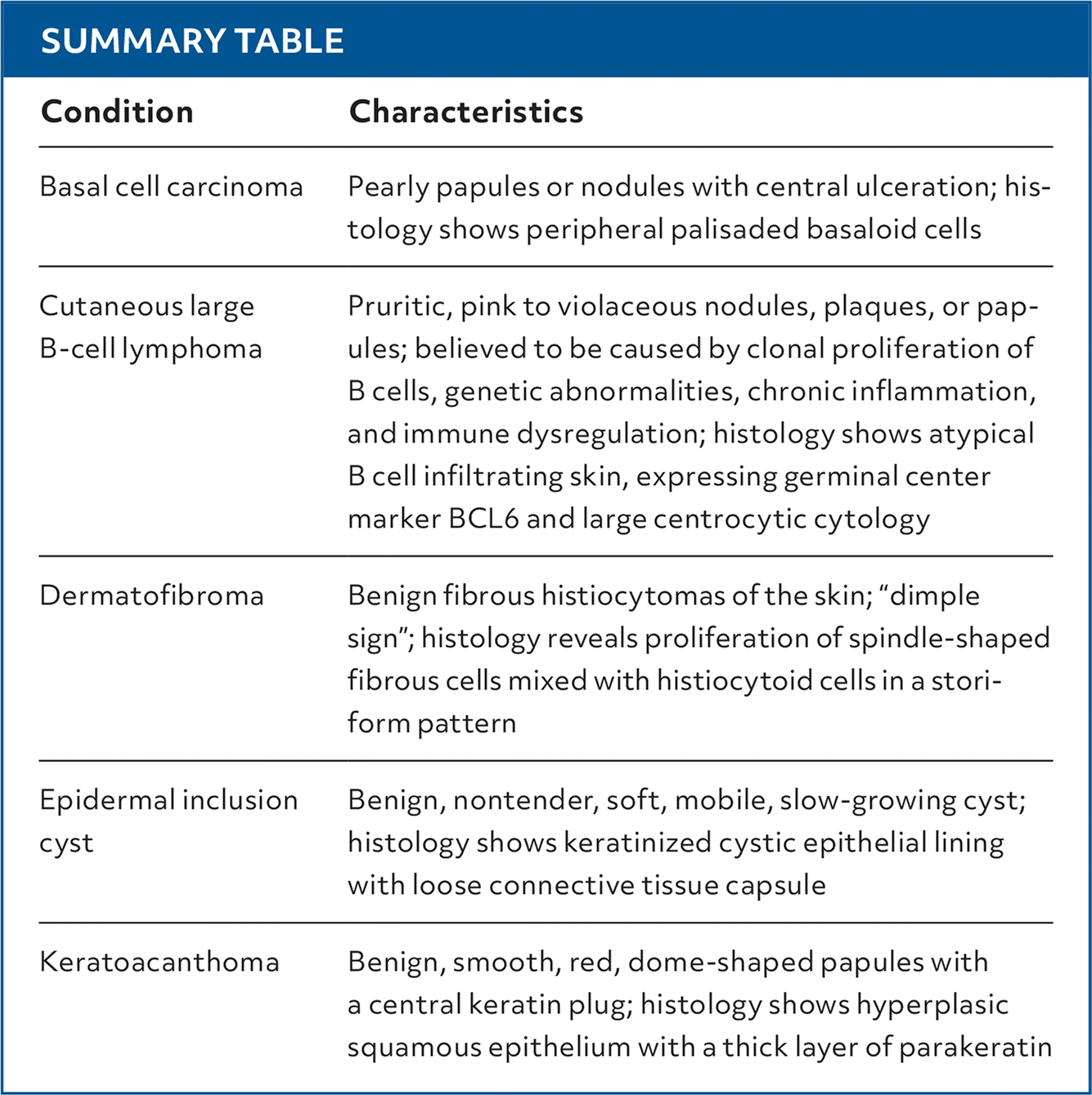
SUMMARY TABLE
| Condition | Characteristics |
|---|---|
| Basal cell carcinoma | Pearly papules or nodules with central ulceration; histology shows peripheral palisaded basaloid cells |
| Cutaneous large B-cell lymphoma | Pruritic, pink to violaceous nodules, plaques, or papules; believed to be caused by clonal proliferation of B cells, genetic abnormalities, chronic inflammation, and immune dysregulation; histology shows atypical B cell infiltrating skin, expressing germinal center marker BCL6 and large centrocytic cytology |
| Dermatofibroma | Benign fibrous histiocytomas of the skin; “dimple sign”; histology reveals proliferation of spindle-shaped fibrous cells mixed with histiocytoid cells in a storiform pattern |
| Epidermal inclusion cyst | Benign, nontender, soft, mobile, slow-growing cyst; histology shows keratinized cystic epithelial lining with loose connective tissue capsule |
| Keratoacanthoma | Benign, smooth, red, dome-shaped papules with a central keratin plug; histology shows hyperplasic squamous epithelium with a thick layer of parakeratin |
Read the full article
Get immediate access, anytime, anywhere.
Choose a single article, issue, or full-access subscription.
Earn up to 14 CME credits per issue.
