

A 41-year-old man presented with a pruritic rash between his toes that had been present for at least 2 months. Treatment with a nonprescription antifungal cream had not led to significant improvement. He did not have any other skin problems, and a review of systems was unremarkable.
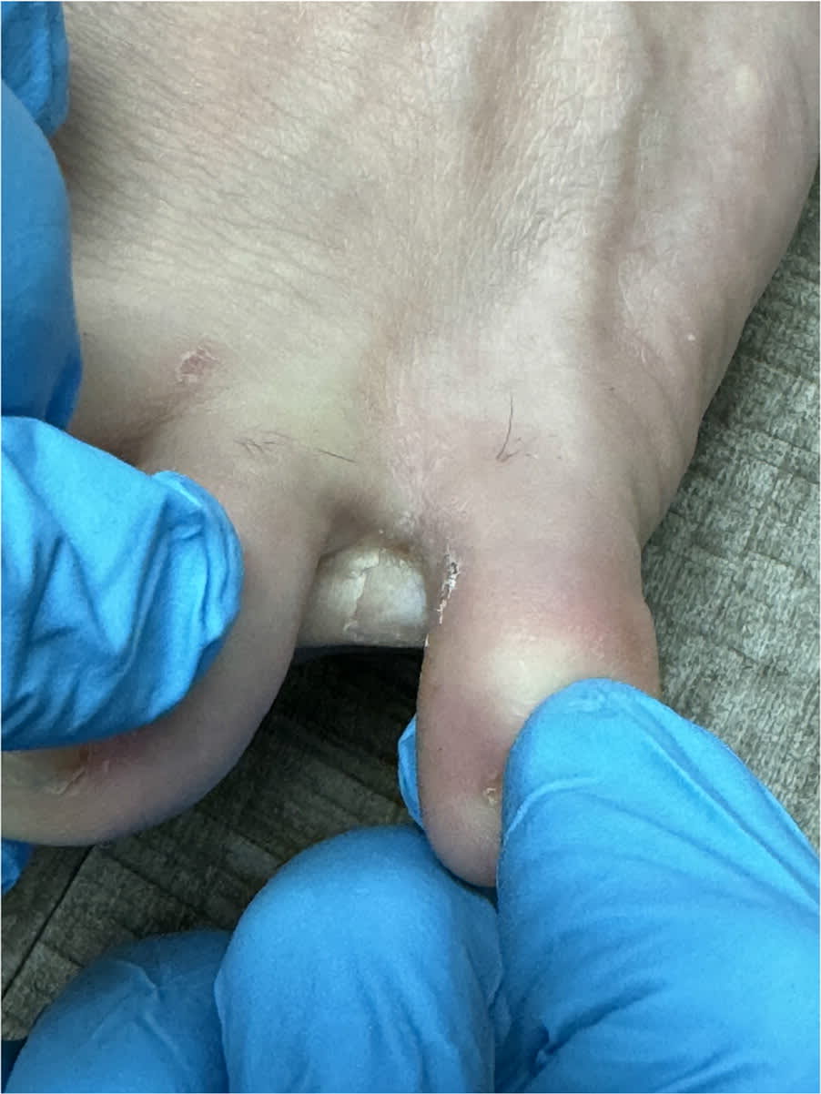
Physical examination revealed moist, macerated, pinkish plaques in the fourth interdigital web spaces bilaterally (Figure 1). Wood lamp examination revealed a distinct coral-red fluorescence.
FIGURE 1
QUESTION
Based on the patient’s history and physical examination, which one of the following is the most likely diagnosis?
- A. Dyshidrotic eczema.
- B. Erythrasma.
- C. Mixed toe web infection.
- D. Psoriasis.
- E. Tinea pedis.
DISCUSSION
The answer is B: erythrasma. This superficial bacterial infection of the skin typically occurs in moist areas such as the interdigital web spaces, groin, axillae, and inframammary fold.1 It is caused by the growth of Corynebacterium minutissimum, gram-positive rods that are part of the natural skin microbiome. Growth is accelerated by heat, moisture, and patient factors, including older age, diabetes, obesity, immunosuppression, hyperhidrosis, and poor hygiene.1,2 Erythrasma comprises up to 17.6% of bacterial skin infections in older patients. The interdigital variety is encountered often in the clinical setting and is the most common bacterial foot infection. C minutissimum cellulitis and bacteremia are rare and occur primarily in immunocompromised individuals.1,2
Dyshidrotic eczema presents as firm, deep-seated vesicles of the lateral and medial aspects of the fingers and less commonly the palms, soles, and toes.1 In some patients, hyperhidrosis is an exacerbating factor. The size of lesions vary. Smaller vesicles can coalesce and create the characteristic look of tapioca pudding.1,3
Erythrasma tends to present as bilateral red-brown hyperpigmented patches with minimal scale in the intertriginous areas.1 When the toes are involved, maceration between the interdigital web spaces is common and may be pruritic. Coral-red fluorescence on Wood lamp examination is confirmatory and due to coproporphyrin III produced by the bacteria. However, fluorescence may not occur if the infected site has been recently cleansed.2
Mixed toe web infections begin with dermatophyte growth, but progressive inflammation and skin damage allow for bacteria growth.1 Patients present with erythematous macerations between digits that can progress to cover the plantar surfaces. Lesions can be purulent, pruritic, and tender to palpation.1,4 The organisms grown from the interdigital spaces include Pseudomonas aeruginosa, Escherichia coli, Proteus mirabilis, and Morganella morganii.4
Psoriasis is an immune system–mediated disorder that presents with inflammatory changes of the epidermis and dermis, resulting in the formation of well-demarcated erythematous plaques with overlying silvery scale on the scalp and extensor surfaces of the elbows, knees, and back.5 Psoriasis can appear anywhere on the body, not just intertriginous areas.1,5
Tinea pedis is a dermatophyte infection of the feet that presents as a pruritic, scaly rash on the soles and may involve the interdigital web spaces.1 It is common in young adults and teenagers.1 No fluorescence occurs with Wood lamp examination. Tinea pedis is diagnosed by presence of septate hyphae on microscopic examination of potassium hydroxide preparation of skin scapings.2
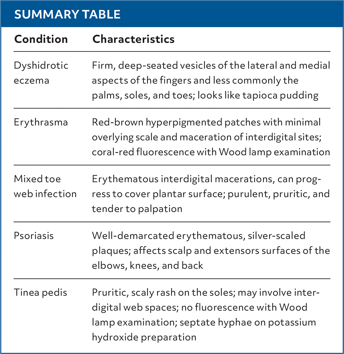
SUMMARY TABLE
| Condition | Characteristics |
|---|---|
| Dyshidrotic eczema | Firm, deep-seated vesicles of the lateral and medial aspects of the fingers and less commonly the palms, soles, and toes; looks like tapioca pudding |
| Erythrasma | Red-brown hyperpigmented patches with minimal overlying scale and maceration of interdigital sites; coral-red fluorescence with Wood lamp examination |
| Mixed toe web infection | Erythematous interdigital macerations, can progress to cover plantar surface; purulent, pruritic, and tender to palpation |
| Psoriasis | Well-demarcated erythematous, silver-scaled plaques; affects scalp and extensors surfaces of the elbows, knees, and back |
| Tinea pedis | Pruritic, scaly rash on the soles; may involve interdigital web spaces; no fluorescence with Wood lamp examination; septate hyphae on potassium hydroxide preparation |
Read the full article
Get immediate access, anytime, anywhere.
Choose a single article, issue, or full-access subscription.
Earn up to 14 CME credits per issue.
