

A 22-year-old man presented with blisters on his lower legs. The lesions were pruritic but not painful. The patient had no other symptoms. On the previous day, he had been in the woods of northern Florida for more than 4 hours in wet conditions with temperatures around 80°F (26.7°C). He was wearing pants and high-ankle boots with socks. He first noticed the blisters that evening.
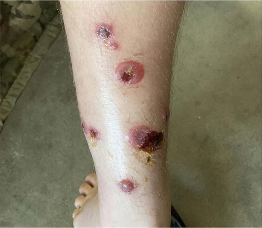
The patient was otherwise healthy and had no significant medical history. He recalled experiencing a similar but milder rash the previous year. Physical examination revealed large bullae on both legs from just above the malleoli to the midcalf area (Figure 1). A few lesions were also found at his waistline. The bullae ruptured and scabbed over 1 week after initial presentation (Figure 2).
FIGURE 1
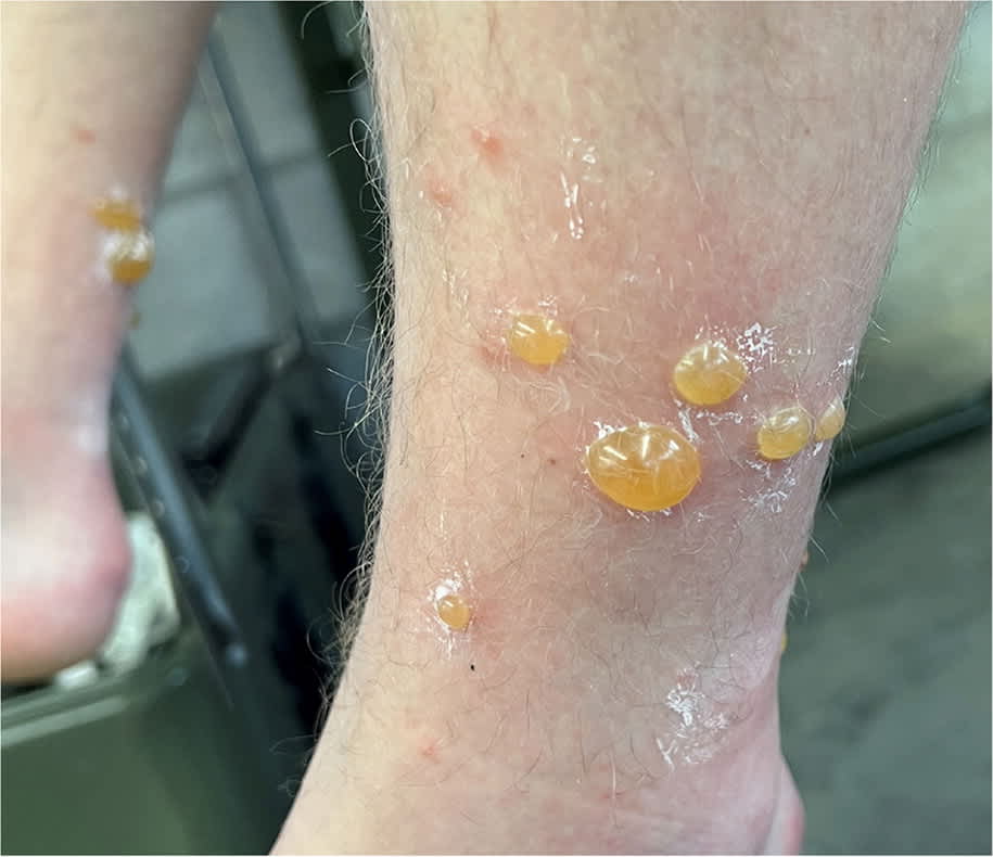
FIGURE 2
QUESTION
Based on the patient’s history and physical examination, which one of the following is the most likely diagnosis?
- A. Bullous hypersensitivity reaction to arthropod bite.
- B. Bullous impetigo.
- C. Bullous pemphigoid.
- D. Bullous reaction to poison ivy.
DISCUSSION
The answer is A: bullous hypersensitivity reaction to arthropod bite.1,2 In this case, Trombiculidae mites (also known as chiggers, red bugs, scrub-itch mites, berry bugs, and harvest mites) are the most likely cause of this uncommon condition. These mites are prevalent in many parts of the United States, including the woods of northern Florida. They typically crawl up shoes and prefer areas where clothing is tight (eg, elastic waistband of underwear, top of socks). Insect repellent containing N, N-diethyl-m-toluamide (DEET) or clothing treated with permethrin can reduce the number of bites.3
Treatment with antihistamines and high-potency topical steroids can reduce pruritus and shorten the duration of the hypersensitivity reaction. Skin hygiene is especially important because a secondary skin infection can occur after the bullae rupture.1
Bullous impetigo is a bacterial skin infection that most commonly occurs in children younger than 2 years. It usually develops within 3 to 5 days of infection and is typically seen on the face. The skin surrounding the bullae is often red and inflamed.
Bullous pemphigoid is most common in adults older than 50 years and often has mucosal involvement. It only occurs in 0.1 to 0.5 per 100,000 patients and is rarely isolated to the lower extremities. It typically includes a long prodromal phase.
A bullous reaction to poison ivy can look similar to arthropod bite hypersensitivity; however, this patient’s history and rash pattern are more consistent with a reaction to an arthropod bite. A reaction to poison ivy generally does not develop in less than 18 hours. It often presents as a linear rash with multiple nonbullous lesions in areas of skin exposure.
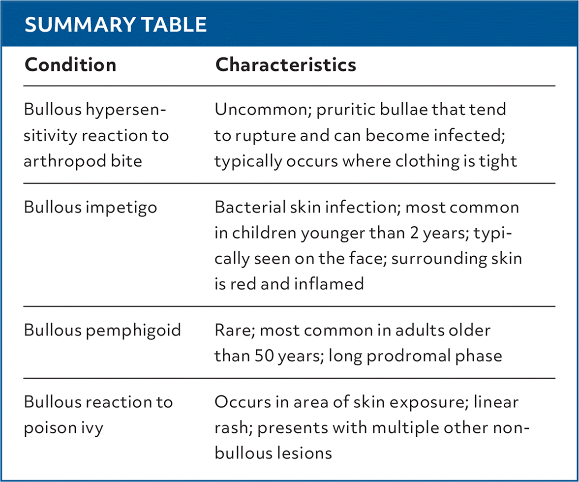
SUMMARY TABLE
| Condition | Characteristics |
|---|---|
| Bullous hypersensitivity reaction to arthropod bite | Uncommon; pruritic bullae that tend to rupture and can become infected; typically occurs where clothing is tight |
| Bullous impetigo | Bacterial skin infection; most common in children younger than 2 years; typically seen on the face; surrounding skin is red and inflamed |
| Bullous pemphigoid | Rare; most common in adults older than 50 years; long prodromal phase |
| Bullous reaction to poison ivy | Occurs in area of skin exposure; linear rash; presents with multiple other non-bullous lesions |
Read the full article
Get immediate access, anytime, anywhere.
Choose a single article, issue, or full-access subscription.
Earn up to 14 CME credits per issue.
