

A 24-hour-old male infant born at 37 weeks of gestation via repeat cesarean delivery was evaluated for emesis that was not bilious or bloody. He had not passed any stools since birth. His prenatal course was complicated by maternal use of buprenorphine to manage an addiction disorder. His mother had declined prenatal genetic testing.
Physical examination revealed slanted palpebral fissures; small, low-set ears; and a flat frontal bridge. He had a large forehead with frontal bossing, short neck with posterior fat pad, and widened space between the first and second toes. Cardiac examination revealed a II/VI holosystolic murmur, best heard at the left lower sternal border. The newborn had a palpable abdominal bowel and a patent anus without meconium plug.
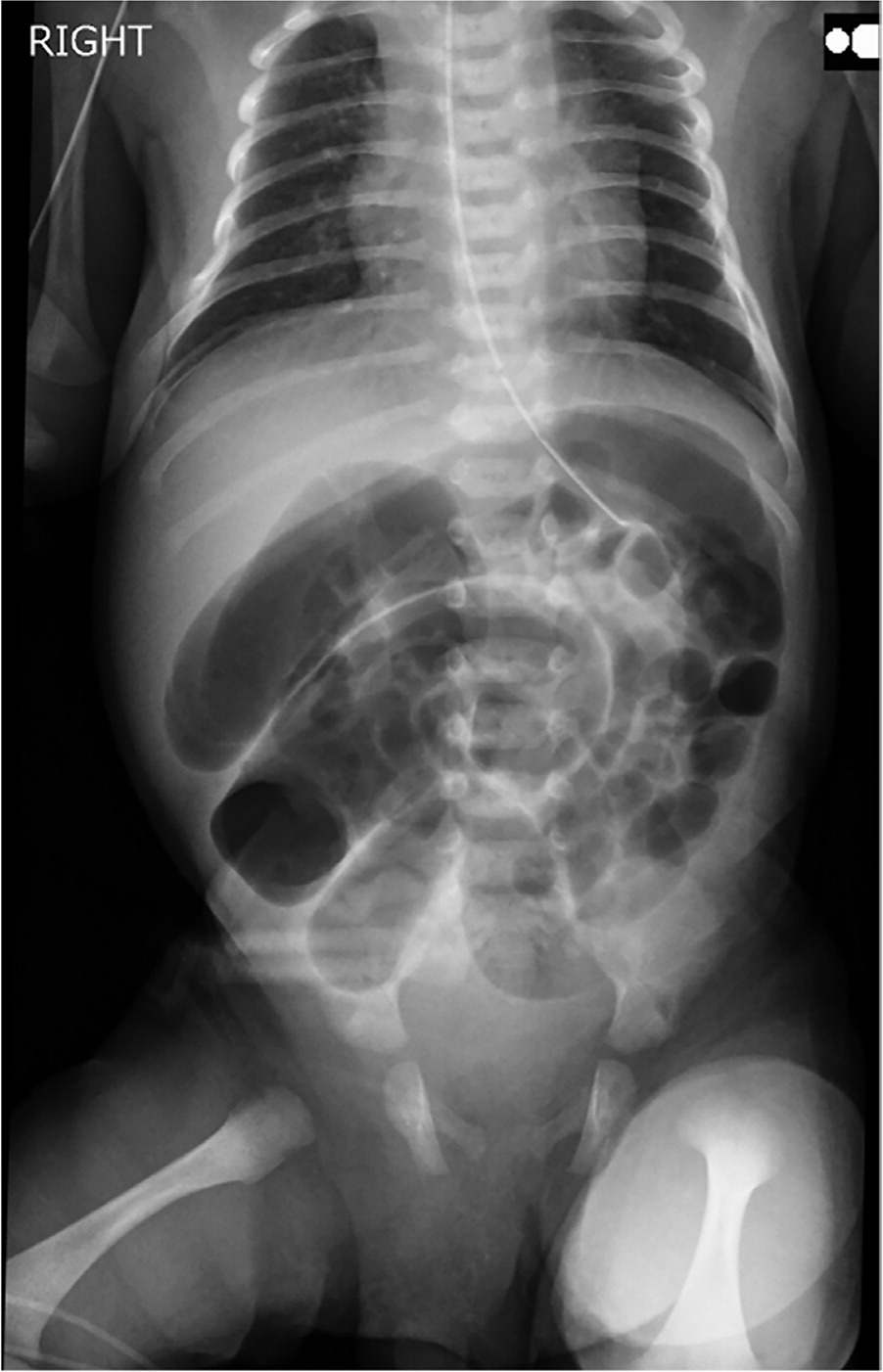
Radiography showed gaseous distention of bowel loops with dilation measuring up to 2.7 cm (Figure 1). No air was identified within the rectum.
FIGURE 1
QUESTION
Based on the patient's history and physical examination, which one of the following is the most likely diagnosis?
- A. Duodenal atresia.
- B. Hirschsprung disease.
- C. Malrotation of the bowel.
- D. Meconium ileus.
- E. Volvulus.
DISCUSSION
The answer is B: Hirschsprung disease, also known as aganglionic megacolon. It occurs in 1 out of 4,000 live births but accounts for 25% of neonatal bowel obstructions.1,2 Hirschsprung disease is four times more likely to occur in male infants and is strongly associated with Down syndrome.1,3 Signs and symptoms include delay or failure to pass meconium with scarce subsequent bowel movements. Obstructive symptoms, including abdominal distention, discomfort, poor oral intake, and vomiting, are common. Diagnosis is based on tissue pathology demonstrating a lack of enteric neurons in the bowel.1 Treatment is surgical removal or bypass of the obstructing colonic segment.
Duodenal atresia is a congenital obstruction in the second portion of the duodenum.3 It is less common than Hirschsprung disease but has a similar predilection for males and association with Down syndrome.3 Diagnosis is clinical, but abdominal radiography may show a characteristic double-bubble sign.3 Treatment is surgical and requires decompression of gastric contents followed by canalization between the second and third portions of the duodenum.
Malrotation is an anatomic abnormality that leads to clockwise twisting of the bowel around the superior mesenteric vessels.3,4 This occurs with incomplete rotation back into the abdominal cavity during fetal development.3 The condition is diagnosed with ultrasonography, which shows malposition of the superior mesenteric vasculature.3 Treatment includes surgical repositioning and bowel excision, depending on the level of mesenteric ischemia.
Meconium ileus accounts for 30% of neonatal bowel obstructions.1 It is caused by increased viscosity of the meconium stool, making it difficult to pass through the bowel and the anus. Meconium ileus is associated with cystic fibrosis.1 Imaging shows enlarged bowel with stool. Treatment includes fluid resuscitation and the use of gastrografin to aid in the passage of stool. In most cases, the bowel is undamaged, but the condition can lead to volvulus, perforation, and peritonitis.
Volvulus is the complete rotation of bowel due to malrotation or narrow, elongated mesentery.3,5,6 Diagnosis is based on imaging showing bowel distention and fluoroscopy showing a characteristic “bird-beak” configuration.3 Treatment is surgical to help prevent mesenteric ischemia.
Read the full article
Get immediate access, anytime, anywhere.
Choose a single article, issue, or full-access subscription.
Earn up to 14 CME credits per issue.
