A 47-year-old woman presented with a solitary raised lesion on her tongue that developed 1 week earlier. Initially, the lesion was red, but it changed to white-pink. It was not painful and did not interfere with chewing, but it occasionally affected her speech. She had no history of trauma, including tongue biting, and no other oral lesions. The patient had no increased sensitivity to hot, cold, or sour stimuli, and she reported no loss or change in taste or other associated symptoms.
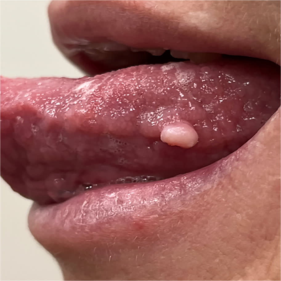
Physical examination revealed a 2- × 3-mm, white-pink papule on the left lateral tongue (Figure 1). It was not tender and did not express pus or fluid. No lymph node swelling was detected.
FIGURE 1
QUESTION
Based on the patient's history and physical examination, which one of the following is the most likely diagnosis?
- A. Giant cell fibroma.
- B. Lymphoepithelial cyst.
- C. Mucocele.
- D. Squamous papilloma.
DISCUSSION
The answer is D: squamous papilloma, which usually occurs as a single, pedunculated or sessile growth of squamous epithelium with papillary projections.1 The surface can appear rounded or cauliflower-like or resemble a finger.2 Squamous papillomas are usually less than 1 cm in size.2 They commonly appear white or red-pink, but the color depends on the level of keratinization, with highly keratinized lesions appearing white.1,2
This benign epithelial neoplasm is caused by human papillomavirus (HPV), most commonly HPV-6 or HPV-11.1 The lesion most often presents in adults 30 to 50 years of age.3 The most common sites are the tongue, lips, palate, or buccal mucosa.1 Squamous papilloma is best diagnosed by biopsy. Histologic features often include exophytic fronds with fibrovascular cores, koilocytes, a stalk, and varying degrees of keratinization.2 Surgical excision is the recommended definitive treatment, and rates of recurrence are low.1
Giant cell fibromas are benign fibrous tumors that can appear in the oral cavity.4 They are often small, pale pink, smooth, dome-shaped nodules that are pedunculated or sessile.4 They are differentiated from other oral lesions when histological examination reveals bi- or multinucleated fibroblasts in the superficial connective tissue, occasionally accompanied by melanin-containing macrophages.4 Giant cell fibromas are not associated with HPV.4 They usually appear in patients 11 to 40 years of age and have a moderate predilection for females.4
Lymphoepithelial cysts most commonly arise in the thyroid, stomach, or pancreas. Oral lymphoepithelial cysts can occur but represent less than 1% of all oral lesions.5 These oral soft-tissue cysts are often yellow to white in color and can appear on the lips, lateral tongue border, hard or soft palate, and cheek mucosa.5 They are slow growing and usually painless but may cause discomfort.6 Excisional biopsy is the preferred treatment, and pathology showing lymphoid follicles in the cyst wall confirms the diagnosis.5
Mucoceles are common in the oral cavity, usually occurring in children and adults younger than 30.7 These lesions result from the rupture of a salivary gland duct. Leakage of mucus into the surrounding soft tissue forms a smooth, translucent vacuole or cyst covered by normal mucosa.8 Oral mucoceles are not related to HPV. They can appear bluish or pink and are usually less than 1 cm in size.7 Oral mucoceles most commonly occur on the lower lip, floor of the mouth, or the inferior surface of the tongue.7
Read the full article
Get immediate access, anytime, anywhere.
Choose a single article, issue, or full-access subscription.
Earn up to 14 CME credits per issue.