Atypical Moles

Liza Straub, MD
Matthew Zeitler, MD
Brian Rayala, MD

American Family Physician. 2026;114(1):27-36.

Author disclosure: No relevant financial relationships.

This clinical content conforms to AAFP criteria for CME.

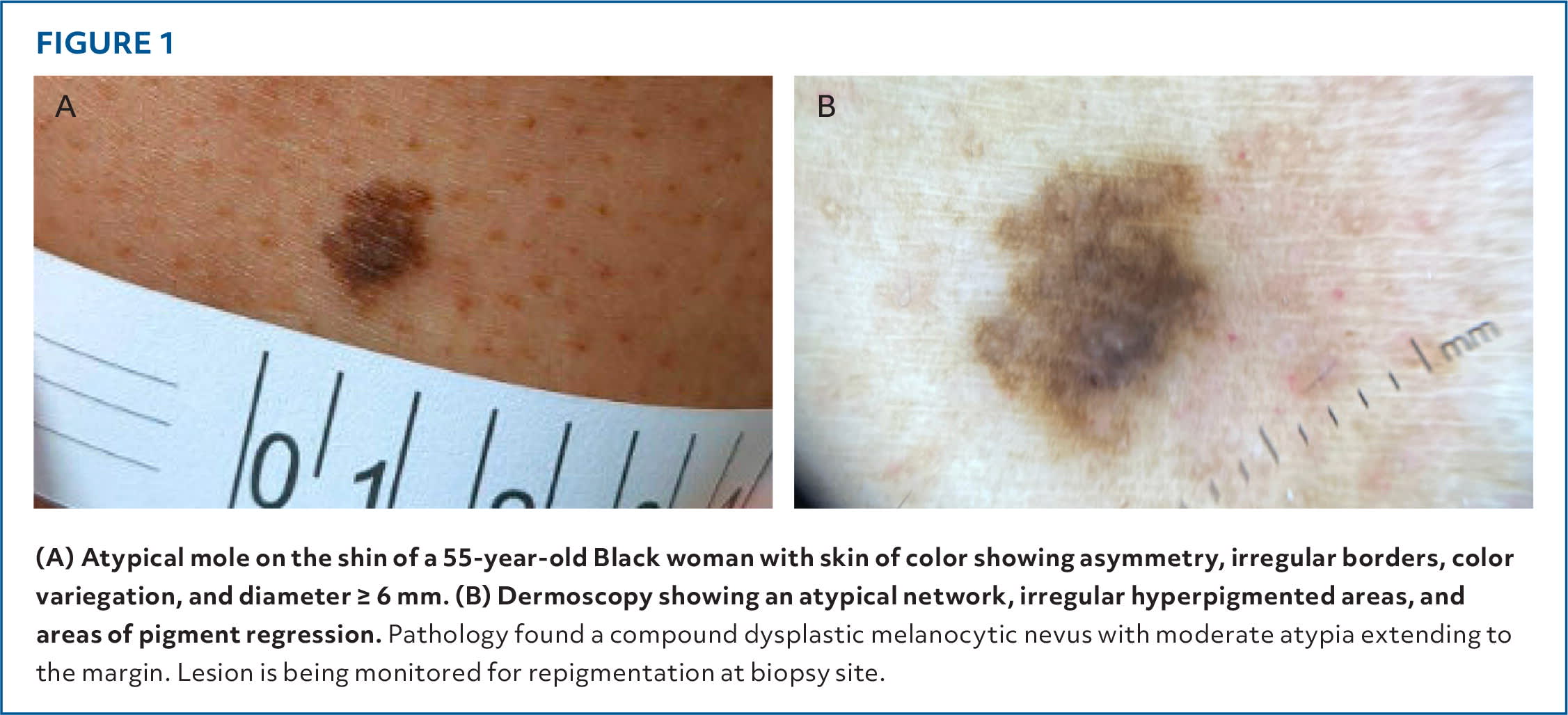
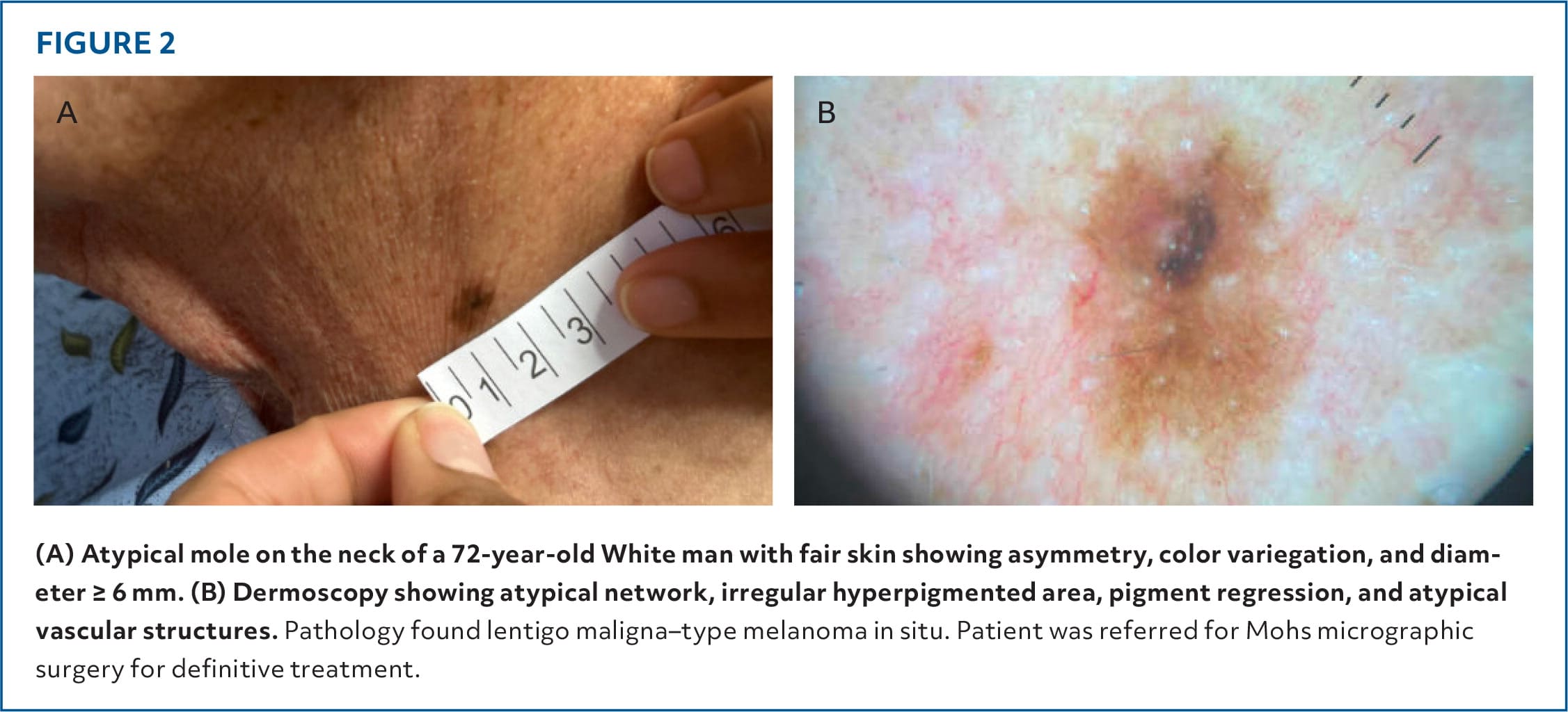
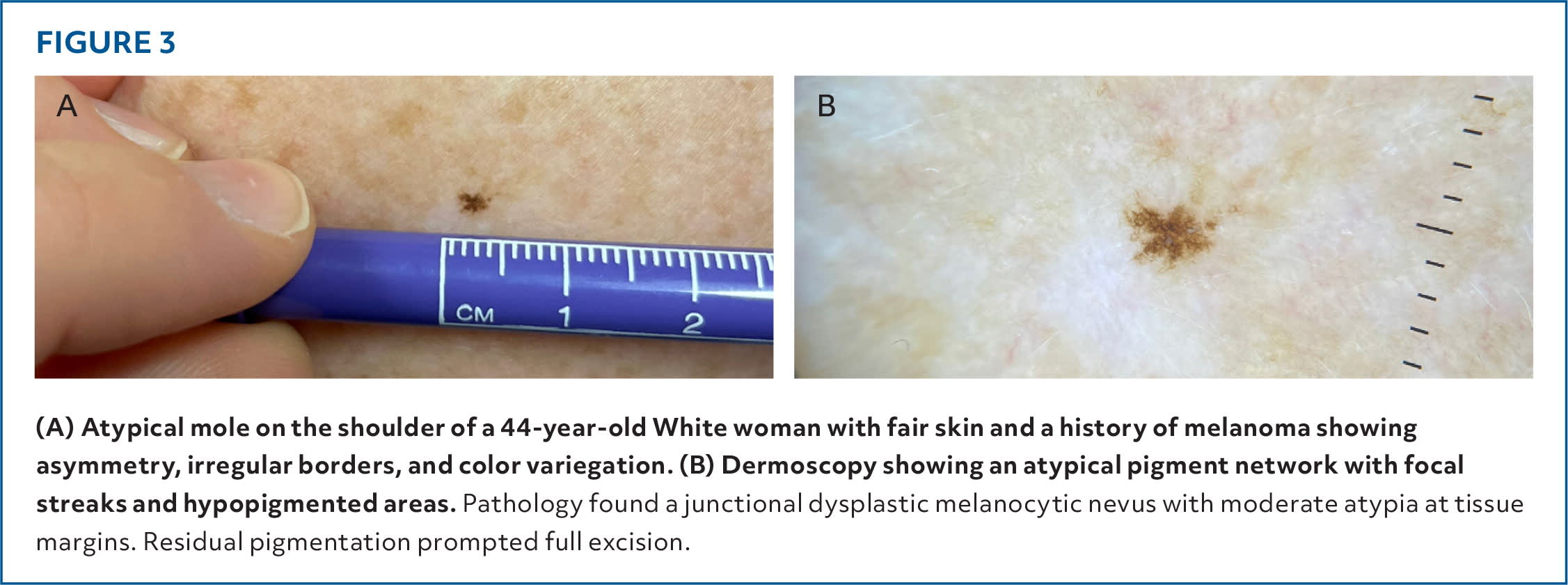
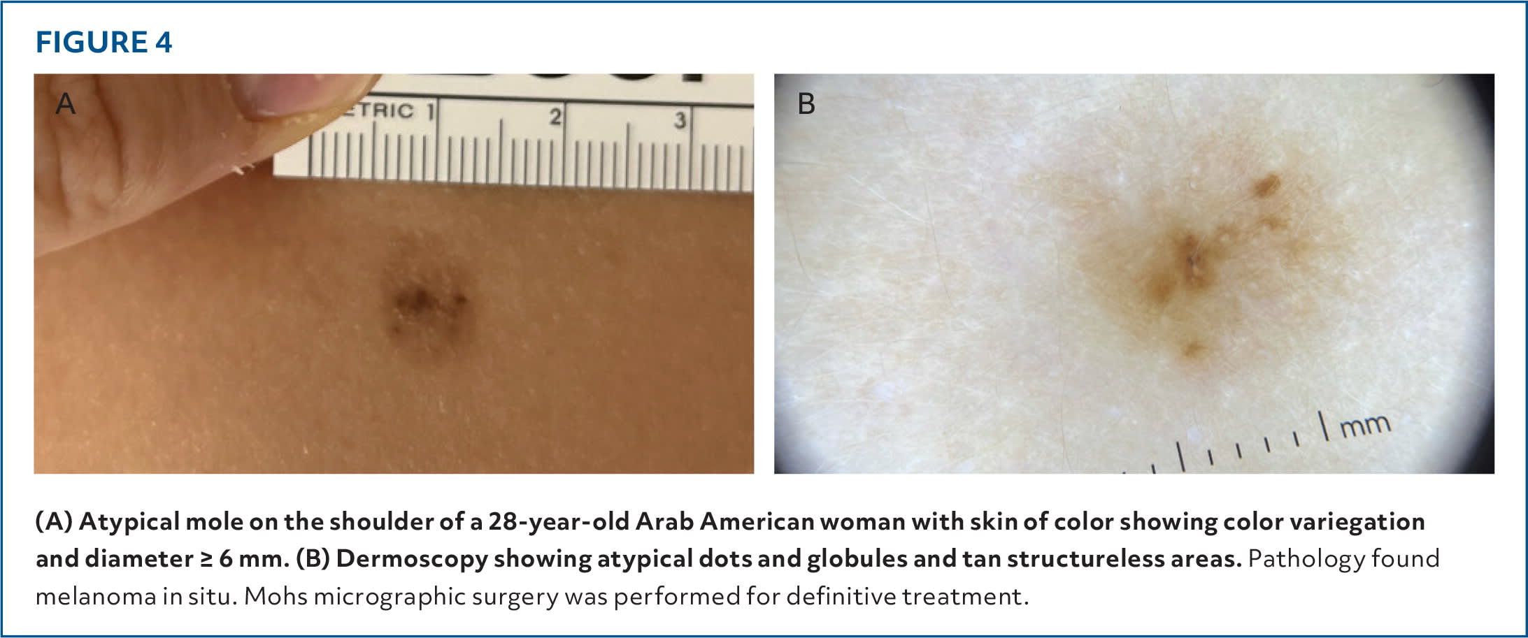
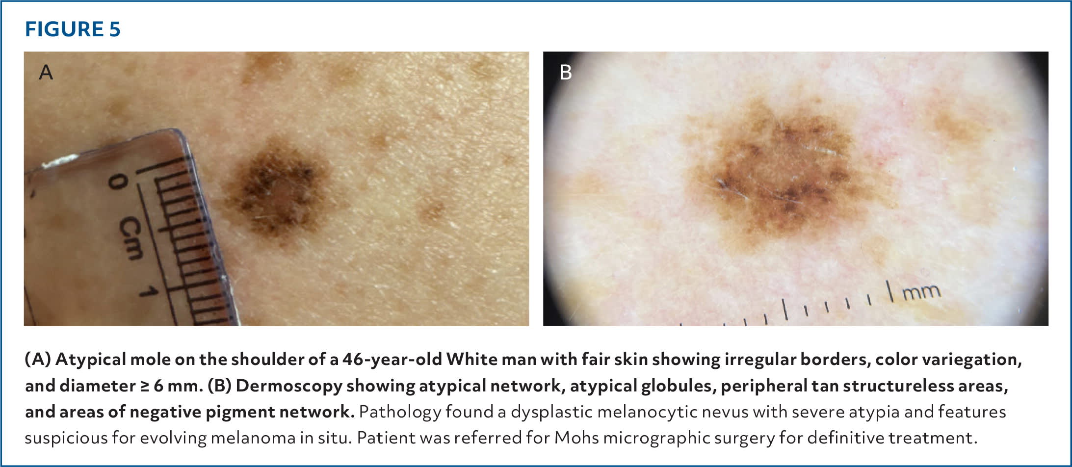
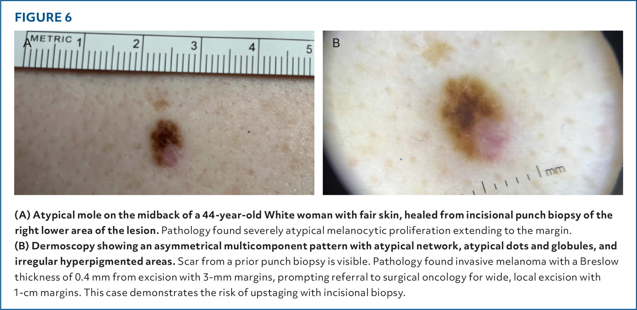
Atypical moles are melanocytic lesions that clinically present with asymmetry, border irregularity, color variegation, or diameter 6 mm or greater. They are more common in patients with fair skin and high cumulative sun exposure. High mole density (more than 100) and presence of atypical moles are associated with increased risk of melanoma and should prompt periodic, systematic skin examinations. The US Preventive Services Task Force recommends patient or parent counseling for individuals with fair skin who are 6 months to 24 years of age on sun protection to reduce skin cancer risk and selective counseling for adults older than 24 years with fair skin and certain risk factors. To distinguish benign moles from melanoma, clinicians should use the ugly duckling assessment and ABCDE (asymmetry, border irregularity, color unevenness, diameter 6 mm or greater, evolution) mnemonic during clinical examination, incorporating dermoscopy if appropriately trained. Biopsy is sometimes necessary to exclude melanoma. Excisional biopsy should include 1- to 3-mm circumferential margins and sufficient depth. Postbiopsy management is guided by the degree of histopathologic atypia determined by the dermatopathologist, margin involvement, and patient risk factors.

LIZA STRAUB, MD, is an assistant professor in the Department of Family Medicine at the University of North Carolina at Chapel Hill School of Medicine, Chapel Hill.

MATTHEW ZEITLER, MD, is an associate professor in the Department of Family Medicine at the University of North Carolina at Chapel Hill School of Medicine, Chapel Hill.

BRIAN RAYALA, MD, is a professor in the Department of Family Medicine at the University of North Carolina at Chapel Hill School of Medicine, Chapel Hill.

Address correspondence to Liza Straub, MD, at liza_straub@med.unc.edu.

Author disclosure: No relevant financial relationships.

  1. 1.Drozdowski R, Spaccarelli N, Peters MS, et al. Dysplastic nevus part I: historical perspective, classification, and epidemiology. J Am Acad Dermatol. 2023;88(1):1-10.
  2. 2.Brunsgaard EK, Wu YP, Grossman D. Melanoma in skin of color: part I. Epidemiology and clinical presentation. J Am Acad Dermatol. 2023;89(3):445-456.
  3. 3.Madankumar R, Gumaste PV, Martires K, et al. Acral melanocytic lesions in the United States: prevalence, awareness, and dermoscopic patterns in skin-of-color and non-Hispanic white patients. J Am Acad Dermatol. 2016;74(4):724.e1-730.e1.
  4. 4.Joshi UM, Kashani-Sabet M, Kirkwood JM. Cutaneous melanoma: a review. JAMA. 2025;334(23):2113-2125.
  5. 5.Weber I, Liao K, Dang T, et al. Sunburn and cutaneous squamous cell carcinoma: a meta-analysis. JAMA Dermatol. 2025;161(11):1148-1156.
  6. 6.Lergenmuller S, Rueegg CS, Perrier F, et al. Lifetime sunburn trajectories and associated risks of cutaneous melanoma and squamous cell carcinoma among a cohort of Norwegian women. JAMA Dermatol. 2022;158(12):1367-1377.
  7. 7.Lashway SG, Worthen ADM, Abuasbeh JN, et al. A meta-analysis of sunburn and basal cell carcinoma risk. Cancer Epidemiol. 2023;85:102379.
  8. 8.Gandini S, Sera F, Cattaruzza MS, et al. Meta-analysis of risk factors for cutaneous melanoma: I. Common and atypical naevi. Eur J Cancer. 2005;41(1):28-44.
  9. 9.El Ghissassi F, Baan R, Straif K, et al.; WHO International Agency for Research on Cancer Monograph Working Group. A review of human carcinogens—part D: radiation. Lancet Oncol. 2009;10(8):751-752.
  10. 10.Gandini S, Sera F, Cattaruzza MS, et al. Meta-analysis of risk factors for cutaneous melanoma: II. Sun exposure. Eur J Cancer. 2005;41(1):45-60.
  11. 11.Pampena R, Kyrgidis A, Lallas A, et al. A meta-analysis of nevus-associated melanoma: prevalence and practical implications. J Am Acad Dermatol. 2017;77(5):938.e4-945.e4.
  12. 12.Pastore LM, Valentini R, Marghoob AA. Congenital melanocytic nevi and risk of melanoma. Clin Dermatol. 2025;43(3):378-384.
  13. 13.Bradford PT, Freedman DM, Goldstein AM, et al. Increased risk of second primary cancers after a diagnosis of melanoma. Arch Dermatol. 2010;146(3):265-272.
  14. 14.Green AC, Olsen CM. Increased risk of melanoma in organ transplant recipients: systematic review and meta-analysis of cohort studies. Acta Derm Venereol. 2015;95(8):923-927.
  15. 15.Gandini S, Sera F, Cattaruzza MS, et al. Meta-analysis of risk factors for cutaneous melanoma: III. Family history, actinic damage and phenotypic factors. Eur J Cancer. 2005;41(14):2040-2059.
  16. 16.Saginala K, Barsouk A, Aluru JS, et al. Epidemiology of melanoma. Med Sci (Basel). 2021;9(4):63.
  17. 17.Eckerle Mize D, Bishop M, Resse E, et al. Familial atypical multiple mole melanoma syndrome. In: Riegert-Johnson DL, Boardman LA, Hefferon T, eds. Cancer Syndromes. National Center for Biotechnology Information; 2009: 67–81.
  18. 18.Grossman DC, Curry SJ, Owens DK, et al. Behavioral counseling to prevent skin cancer: US Preventive Services Task Force recommendation statement. JAMA. 2018;319(11):1134-1142.
  19. 19.Wehner MR. Sunscreen and melanoma prevention: evidence and expectations. Br J Dermatol. 2018;178(1):15-16.
  20. 20.Olsen CM, Wilson LF, Green AC, et al. How many melanomas might be prevented if more people applied sunscreen regularly?. Br J Dermatol. 2018;178(1):140-147.
  21. 21.American Academy of Dermatology. Sunscreen FAQs. Updated February 11, 2025. Accessed June 11, 2025. https://www.aad.org/media/stats-sunscreen
  22. 22.Centers for Disease Control and Prevention. Sun safety facts. February 10, 2026. Accessed June 9, 2026. https://www.cdc.gov/skin-cancer/sun-safety/index.html
  23. 23.Sohn M, Herzog B, Osterwalder U, et al. Calculation of the sun protection factor of sunscreens with different vehicles using measured film thickness distribution—comparison with the SPF in vitro. J Photochem Photobiol B. 2016;159:74-81.
  24. 24.Williams JD, Maitra P, Atillasoy E, et al. SPF 100+ sunscreen is more protective against sunburn than SPF 50+ in actual use: results of a randomized, double-blind, split-face, natural sunlight exposure clinical trial. J Am Acad Dermatol. 2018;78(5):902.e2-910.e2.
  25. 25.Kohli I, Nicholson CL, Williams JD, et al. Greater efficacy of SPF 100+ sunscreen compared with SPF 50+ in sunburn prevention during 5 consecutive days of sunlight exposure: a randomized, double-blind clinical trial. J Am Acad Dermatol. 2020;82(4):869-877.
  26. 26.Solish N, Humphrey S, Waller B, et al. Photoprotection with mineral-based sunscreens. Dermatol Surg. 2020;46(12):1508-1513.
  27. 27.Centers for Disease Control and Prevention. Sun exposure in travelers. April 23, 2025. Accessed June 11, 2025. https://www.cdc.gov/yellow-book/hcp/environmental-hazards-risks/sun-exposure-in-travelers.html
  28. 28.Weiss BD. Should your patients avoid sunscreen? [editorial]. Am Fam Physician. 2024;110(6):566-567.
  29. 29.Perez M, Abisaad JA, Rojas KD, et al. Skin cancer: primary, secondary, and tertiary prevention. Part I.. J Am Acad Dermatol. 2022;87(2):255-268.
  30. 30.Centers for Disease Control and Prevention. Public health image library. ID# 18405. Accessed June 9, 2026. https://phil.cdc.gov/Details.aspx?pid=18405
  31. 31.Abbasi NR, Shaw HM, Rigel DS, et al. Early diagnosis of cutaneous melanoma: revisiting the ABCD criteria. JAMA. 2004;292(22):2771-2776.
  32. 32.Rigel DS, Russak J, Friedman R. The evolution of melanoma diagnosis: 25 years beyond the ABCDs. CA Cancer J Clin. 2010;60(5):301-316.
  33. 33.Tsao H, Olazagasti JM, Cordoro KM, et al.; American Academy of Dermatology Ad Hoc Task Force for the ABCDEs of Melanoma. Early detection of melanoma: reviewing the ABCDEs. J Am Acad Dermatol. 2015;72(4):717-723.
  34. 34.Ilyas M, Costello CM, Zhang N, et al. The role of the ugly duckling sign in patient education. J Am Acad Dermatol. 2017;77(6):1088-1095.
  35. 35.Chen JY, Fernandez K, Fadadu RP, et al. Skin cancer diagnosis by lesion, physician, and examination type: a systematic review and meta-analysis. JAMA Dermatol. 2025;161(2):135-146.
  36. 36.Dinnes J, Deeks JJ, Chuchu N, et al.; Cochrane Skin Cancer Diagnostic Test Accuracy Group. Dermoscopy, with and without visual inspection, for diagnosing melanoma in adults. Cochrane Database Syst Rev. 2018(12):CD011902.
  37. 37.Perkins A, Duffy RL. Atypical moles: diagnosis and management. Am Fam Physician. 2015;91(11):762-767.
  38. 38.Marghoob AA, Usatine RP, Jaimes N. Dermoscopy for the family physician. Am Fam Physician. 2013;88(7):441-450.
  39. 39.Argenziano G, Catricalà C, Ardigo M, et al. Seven-point checklist of dermoscopy revisited. Br J Dermatol. 2011;164(4):785-790.
  40. 40.Dolianitis C, Kelly J, Wolfe R, et al. Comparative performance of 4 dermoscopic algorithms by nonexperts for the diagnosis of melanocytic lesions. Arch Dermatol. 2005;141(8):1008-1014.
  41. 41.Rosendahl C, Tschandl P, Cameron A, et al. Diagnostic accuracy of dermatoscopy for melanocytic and nonmelanocytic pigmented lesions. J Am Acad Dermatol. 2011;64(6):1068-1073.
  42. 42.Mangione CM, Barry MJ, Nicholson WK, et al. Screening for skin cancer: US Preventive Services Task Force recommendation statement. JAMA. 2023;329(15):1290-1295.
  43. 43.American Academy of Family Physicians. Cancer care: clinical guidance and practice resources: skin cancer. Accessed May 27, 2026. https://www.aafp.org/clinical-insights/preventive-care-and-whole-health/cancer-care
  44. 44.Rojas KD, Perez ME, Marchetti MA, et al. Skin cancer: primary, secondary, and tertiary prevention. Part II. J Am Acad Dermatol. 2022;87(2):271-288.
  45. 45.Naeyaert JM, Brochez L. Clinical practice. Dysplastic nevi. N Engl J Med. 2003;349(23):2233-2240.
  46. 46.Fried L, Tan A, Bajaj S, et al. Technological advances for the detection of melanoma: advances in diagnostic techniques. J Am Acad Dermatol. 2020;83(4):983-992.
  47. 47.Swetter SM, Tsao H, Bichakjian CK, et al. Guidelines of care for the management of primary cutaneous melanoma. J Am Acad Dermatol. 2019;80(1):208-250.
  48. 48.Lauters R, Brown AD, Harrington KCA. Melanoma: diagnosis and treatment. Am Fam Physician. 2024;110(4):367-377.
  49. 49.Michielin O, van Akkooi ACJ, Ascierto PA, et al. Cutaneous melanoma: ESMO clinical practice guidelines for diagnosis, treatment and follow-up. Ann Oncol. 2019;30(12):1884-1901.
  50. 50.Pathak S, Puckett Y. Cutaneous malignant melanoma: guideline-based management and interprofessional collaboration. StatPearls. Updated June 29, 2025. Accessed June 9, 2026. https://www.ncbi.nlm.nih.gov/books/NBK572149
  51. 51.Chacko RS, Shah PC, Feng JX, et al. Risk of tumor upstaging following partial biopsy and scouting biopsy in the treatment of melanoma in situ and invasive melanoma with Mohs micrographic surgery: a retrospective cohort study. Arch Dermatol Res. 2025;317(1):659.
  52. 52.Tran KT, Wright NA, Cockerell CJ. Biopsy of the pigmented lesion— when and how. J Am Acad Dermatol. 2008;59(5):852-871.
  53. 53.Kim CC, Swetter SM, Curiel-Lewandrowski C, et al. Addressing the knowledge gap in clinical recommendations for management and complete excision of clinically atypical nevi/dysplastic nevi: Pigmented Lesion Subcommittee consensus statement. JAMA Dermatol. 2015;151(2):212-218.
  54. 54.Pickett H. Shave and punch biopsy for skin lesions. Am Fam Physician. 2011;84(9):995-1002.
  55. 55.Etzkorn JR, Sharkey JM, Grunyk JW, et al. Frequency of and risk factors for tumor upstaging after wide local excision of primary cutaneous melanoma. J Am Acad Dermatol. 2017;77(2):341-348.
  56. 56.Hocker TL, Alikhan A, Comfere NI, et al. Favorable long-term outcomes in patients with histologically dysplastic nevi that approach a specimen border. J Am Acad Dermatol. 2013;68(4):545-551.
  57. 57.Spaccarelli N, Drozdowski R, Peters MS, et al. Dysplastic nevus part II: dysplastic nevi: molecular/genetic profiles and management. J Am Acad Dermatol. 2023;88(1):13-20.
  58. 58.Maghari A. Dysplastic (or atypical) nevi showing moderate or severe atypia with clear margins on the shave removal specimens are most likely completely excised. J Cutan Med Surg. 2017;21(1):42-47.
  59. 59.Hiscox B, Hardin MR, Orengo IF, et al. Recurrence of moderately dysplastic nevi with positive histologic margins. J Am Acad Dermatol. 2017;76(3):527-530.
  60. 60.Kim CC, Berry EG, Marchetti MA, et al.; Pigmented Lesion Subcommittee, Melanoma Prevention Working Group. Risk of subsequent cutaneous melanoma in moderately dysplastic nevi excisionally biopsied but with positive histologic margins. JAMA Dermatol. 2018;154(12):1401-1408.
  61. 61.Reddy KK, Farber MJ, Bhawan J, et al. Atypical (dysplastic) nevi: outcomes of surgical excision and association with melanoma. JAMA Dermatol. 2013;149(8):928-934.
  62. 62.Engeln K, Peters K, Ho J, et al. Dysplastic nevi with severe atypia: long-term outcomes in patients with and without re-excision. J Am Acad Dermatol. 2017;76(2):244-249.
  63. 63.Blum A, Hofmann-Wellenhof R, Marghoob AA, et al. Recurrent melanocytic nevi and melanomas in dermoscopy: results of a multicenter study of the International Dermoscopy Society. JAMA Dermatol. 2014;150(2):138-145.
  64. 64.Soleymani T, Swetter SM, Hollmig ST, et al. Adequacy of conservative 2- to 3-mm surgical margins for complete excision of biopsy-proven severely dysplastic nevi: retrospective case series at a tertiary academic institution. J Am Acad Dermatol. 2020;83(1):254-255.
  65. 65.Fleming NH, Shaub AR, Bailey E, et al. Outcomes of surgical re-excision versus observation of severely dysplastic nevi: a single-institution, retrospective cohort study. J Am Acad Dermatol. 2020;82(1):238-240.
  66. 66.Cyr PR. Atypical moles. Am Fam Physician. 2008;78(6):735-740.

Copyright © 2026 by the American Academy of Family Physicians.

This content is owned by the AAFP. A person viewing it online may make one printout of the material and may use that printout only for his or her personal, non-commercial reference. This material may not otherwise be downloaded, copied, printed, stored, transmitted or reproduced in any medium, whether now known or later invented, except as authorized in writing by the AAFP. See permissions for copyright questions and/or permission requests.