Diabetic ulcers are the most common foot injuries leading to lower extremity amputation. Family physicians have a pivotal role in the prevention or early diagnosis of diabetic foot complications. Management of the diabetic foot requires a thorough knowledge of the major risk factors for amputation, frequent routine evaluation and meticulous preventive maintenance. The most common risk factors for ulcer formation include diabetic neuropathy, structural foot deformity and peripheral arterial occlusive disease. A careful physical examination, buttressed by monofilament testing for neuropathy and noninvasive testing for arterial insufficiency, can identify patients at risk for foot ulcers and appropriately classify patients who already have ulcers or other diabetic foot complications. Patient education regarding foot hygiene, nail care and proper footwear is crucial to reducing the risk of an injury that can lead to ulcer formation. Adherence to a systematic regimen of diagnosis and classification can improve communication between family physicians and diabetes subspecialists and facilitate appropriate treatment of complications. This team approach may ultimately lead to a reduction in lower extremity amputations related to diabetes.
Diabetic foot complications are the most common cause of nontraumatic lower extremity amputations in the industrialized world. The risk of lower extremity amputation is 15 to 46 times higher in diabetics than in persons who do not have diabetes mellitus.1,2 Furthermore, foot complications are the most frequent reason for hospitalization in patients with diabetes, accounting for up to 25 percent of all diabetic admissions in the United States and Great Britain.3–5
The vast majority of diabetic foot complications resulting in amputation begin with the formation of skin ulcers. Early detection and appropriate treatment of these ulcers may prevent up to 85 percent of amputations.6,7 Indeed, one of the disease prevention objectives outlined in the “Healthy People 2000” project of the U.S. Department of Health and Human Services is a 40 percent reduction in the amputation rate for diabetic patients. Family physicians have an integral role in ensuring that patients with diabetes receive early and optimal care for skin ulcers.
Unfortunately, several studies8,9 have found that primary care physicians infrequently perform foot examinations in diabetic patients during routine office visits. The feet of hospitalized diabetics may also be inadequately evaluated.10
Careful inspection of the diabetic foot on a regular basis is one of the easiest, least expensive and most effective measures for preventing foot complications. Appropriate care of the diabetic foot requires recognition of the most common risk factors for limb loss. Many of these risk factors can be identified based on specific aspects of the history and a brief but systematic examination of the foot.
Risk Factors for Lower Extremity Amputation
Common risk factors for amputation of the diabetic foot include peripheral neuropathy, structural foot deformity, ulceration, infection and peripheral vascular disease4 (Table 1). It is important to recognize that foot ulcers can have a multifactorial etiology.
TABLE 1 Risk Factors for Lower Extremity Amputation in the Diabetic Foot
| Absence of protective sensation due to peripheral neuropathy |
| Arterial insufficiency |
| Foot deformity and callus formation resulting in focal areas of high pressure |
| Autonomic neuropathy causing decreased sweating and dry, fissured skin |
| Limited joint mobility |
| Obesity |
| Impaired vision |
| Poor glucose control leading to impaired wound healing |
| Poor footwear that causes skin breakdown or inadequately protects the skin from high pressure and shear forces |
| History of foot ulcer or lower extremity amputation |
Peripheral Arterial Occlusive Disease
Peripheral arterial occlusive disease is four times more prevalent in diabetics than in non-diabetics.11 The arterial occlusion typically involves the tibial and peroneal arteries but spares the dorsalis pedis artery.12 Smoking, hypertension and hyperlipidemia commonly contribute to the increased prevalence of peripheral arterial occlusive disease in diabetics.13,14
The presence of lower extremity ischemia is suggested by a combination of clinical signs and symptoms plus abnormal results on noninvasive vascular tests. Signs and symptoms may include claudication, pain occurring in the arch or forefoot at rest or during the night, absent popliteal or posterior tibial pulses, thinned or shiny skin, absence of hair on the lower leg and foot, thickened nails, redness of the affected area when the legs are dependent, or “dangled,” and pallor when the foot is elevated.
Noninvasive vascular tests include transcutaneous oxygen measurement,15 the ankle-brachial index (ABI) and the absolute toe systolic pressure.16,17 The ABI is a noninvasive test that can be performed easily in the office using a handheld Doppler device. A blood pressure cuff is placed on the upper arm and inflated until no brachial pulse is detected by the Doppler device. The cuff is then slowly deflated until a Doppler-detected pulse returns (the systolic pressure). This maneuver is repeated on the leg, with the cuff wrapped around the distal calf and the Doppler device placed over the dorsalis pedis or posterior tibial artery. The ankle systolic pressure divided by the brachial systolic pressure gives the ABI.
The sensitivity and specificity of noninvasive vascular tests are a matter of some controversy. Commonly accepted abnormal values for transcutaneous oxygen measurement, ABI determinations and toe systolic pressure are given in Table 2. The noninvasive tests have been faulted for underestimating the severity of arterial insufficiency.18 If lower extremity ischemia is strongly suspected, arteriography or some other imaging study should be performed to confirm or rule out ischemia.
TABLE 2 Noninvasive Vascular Tests
| Test | Abnormal value |
|---|---|
| Transcutaneous oxygen measurement | Less than 40 mm Hg |
| Ankle-brachial index | Less than 0.80: abnormal |
| Less than 0.45: severe, limb-threatening | |
| Absolute toe systolic pressure | Less than 45 mm Hg |
Optimal ulcer healing requires adequate tissue perfusion. Thus, arterial insufficiency should be suspected if an ulcer fails to heal. Vascular surgery consultation and possible revascularization should be considered when clinical signs of ischemia are present in the lower extremity of a diabetic patient and the results of noninvasive vascular tests or imaging studies suggest that the patient has peripheral arterial occlusive disease.
Proper control of concomitant hypertension or hyperlipidemia can help to reduce the risk of peripheral arterial occlusive disease. Smoking cessation is essential for preventing the progression of occlusive disease.
Sensory and Autonomic Neuropathy
Distal symmetric polyneuropathy is perhaps the most common complication affecting the lower extremities of patients with diabetes mellitus. This complication occurs in up to 58 percent of patients with longstanding disease.19 Neuropathy, a major etiologic component of most diabetic ulcerations, is present in more than 82 percent of diabetic patients with foot wounds.4 This lack of protective sensation, combined with unaccommodated foot deformities, exposes patients to undue sudden or repetitive stress that leads to eventual ulcer formation with a risk of infection and possible amputation.20
In the diabetic foot, autonomic neuropathy has several common manifestations. First, denervation of dermal structures leads to decreased sweating. This causes dry skin and fissure formation, which predispose the skin to infection. In vascularly competent patients, this “autosympathectomy” may lead to increased blood flow, which has been implicated as one of the primary etiologic factors in the development of Charcot's joint and severe foot deformity.21–23
The nylon monofilament test is a simply performed office test to diagnose patients at risk for ulcer formation due to peripheral sensory neuropathy.24 The test is abnormal if the patient cannot sense the touch of the monofilament when it is pressed against the foot with just enough pressure to bend the filament25 (Figure 1). Physicians can obtain a monofilament kit (and literature on diabetic foot management) at a small cost from the National Diabetes Information Clearing-house (301-654-3327).
FIGURE 1.
Nylon monofilament test. There is a risk of ulcer formation if the patient is unable to feel the monofilament when it is pressed against the foot with just enough pressure to bend the filament. The patient is asked to say “yes” each time he or she feels the filament. Failure to feel the filament at four of 10 sites is 97 percent sensitive and 83 percent specific for identifying loss of protective sensation.
Structural Deformity and Limited Joint Mobility
Foot deformities, which are common in diabetic patients, lead to focal areas of high pressure. When an abnormal focus of pressure is coupled with lack of sensation, a foot ulcer can develop. Most diabetic foot ulcers form over areas of bony prominences (Figure 2), especially when bunions, calluses or hammer-toe formations lead to abnormally prominent bony points. Foot deformities are believed to be more common in diabetic patients due to atrophy of the intrinsic musculature responsible for stabilizing the toes.20
FIGURE 2.
Usual locations of ulcers in the diabetic foot. Ulceration is particularly likely to occur over the dorsal portion of the toes and on the plantar aspect of the metatarsal heads and the heel.
Rigid deformities or limited range of motion at the subtalar or metatarsophalangeal joints have also been associated with the development of diabetic foot ulcers.26,27 Other mechanisms of skin breakdown in the insensate diabetic foot include puncture wounds and thermal injuries from, for example, hot water soaks.
History of Previous Ulceration and Amputation
A diabetic patient with a history of previous ulceration or amputation is at increased risk for further ulceration, infection and subsequent amputation. Alterations in foot dynamics due to ulceration, joint deformity or amputation can cause the abnormal distribution of plantar pressures and result in the formation of new ulcers28 (Figure 3).
Figure 3.
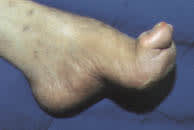
Structural deformity. When combined with sensory neuropathy, a structural foot deformity may predispose the diabetic patient to ulceration, infection and subsequent amputation.
Prevention of Ulcer Formation
Meticulous attention to foot care and proper management of minor foot injuries are key to preventing ulcer formation. Daily foot inspection by the patient (or a caretaker if the patient lacks sufficient visual acuity or mobility to perform the examination) is the cornerstone of proper foot care. Gentle cleansing with soap and water, followed by the application of topical moisturizers, helps to maintain healthy skin that can better resist breakdown and injury.
The physician should inspect the patient's shoes for areas of inadequate support or improper fit. While many patients do well with commercially available athletic shoes and thick, absorbent socks, patients with foot deformities or special support needs may benefit from custom shoes. Medicare Part B now covers the purchase of custom shoes when the certifying physician identifies a risk factor for ulcer formation and submits appropriate documentation. A sample documentation form is provided with the monofilament kit used to test patients for peripheral sensory neuropathy.
Minor foot injuries and infections, such as cuts, scrapes, blisters and tinea pedis, can be unintentionally exacerbated by home remedies that impede healing. Patients should be reminded to avoid hot soaks, heating pads and harsh topical agents such as hydrogen peroxide, iodine (e.g., Betadine) and astringents (e.g., witch hazel). Gentle cleansing of minor wounds and the application of a topical antibiotic to maintain a moist wound environment can help to prevent ulcer formation. In addition, the physician should inspect any minor wound that does not heal rapidly.
By reinforcing preventive advice and inspecting the patient's feet at routine follow-up visits, the physician can help the patient develop and maintain good foot-care habits.
Ulceration
Despite the best intentions and careful attention to foot care, many diabetic patients eventually develop foot ulcers. These wounds are the principal portal of entry for infection in patients with diabetes (Figure 4). Frequently, the ulcers are covered by callus or fibrotic tissue. This makes the trimming of hyperkeratotic tissue important for comprehensive wound evaluation.
Figure 4.
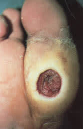
Neuropathic ulceration of the foot in a diabetic patient.
Because these ulcers almost always form in patients with neuropathy, they are typically painless. Even in the presence of severe infection, many patients have few subjective complaints and are often more concerned with soiled footwear and stockings than with the penetrating wound.29
Adequate debridement is the first step in the evaluation of a foot ulcer. Debridement should remove all necrotic tissue and surrounding callus until a healthy bleeding edge is revealed. Patients (and physicians) often underestimate the need for debridement and may be surprised by the appearance of the newly debrided ulcer.18 Topical debriding enzymes are expensive and have not been conclusively shown to be beneficial.
After debridement, the ulcer should be probed with a sterile blunt instrument to determine the involvement of underlying structures, such as tendon, joint capsule or bone. Probing to bone is a simple and specific test for osteomyelitis, but it has low sensitivity.30 Plain-film radiographs should be obtained to look for soft tissue gas and foreign bodies and to evaluate the ulcer for bone involvement.
It can be difficult to differentiate local soft tissue infection and inflammation from osteomyelitis. Three-phase bone scans and radiolabelled leukocyte scans are expensive but can help to establish an accurate diagnosis in problematic cases.31 The involvement of underlying structures and the presence or absence of ischemia and/or infection must be determined before an appropriate wound classification can be made and a subsequent treatment plan can be implemented.32
A variety of wound classification systems exists. Table 333,34 outlines one diabetic foot classification system that is presently being evaluated to determine if its use will reduce the incidence of diabetic foot amputations. This classification system divides the findings for the diabetic foot into six categories based on increasing risk. The first three categories are risk factors for foot ulceration, and the second three are risk factors for amputation. The suggested treatments reflect the degree of risk for each category.
Recognition of risk factors, preventive foot maintenance and regular foot examinations are essential in preventing foot ulcers in patients with diabetes. When foot ulcers develop despite preventive measures, a systematically applied regimen of diagnosis and classification, coupled with early and appropriate treatment, should help to reduce the tremendous personal and societal burden of diabetes-related amputations.
