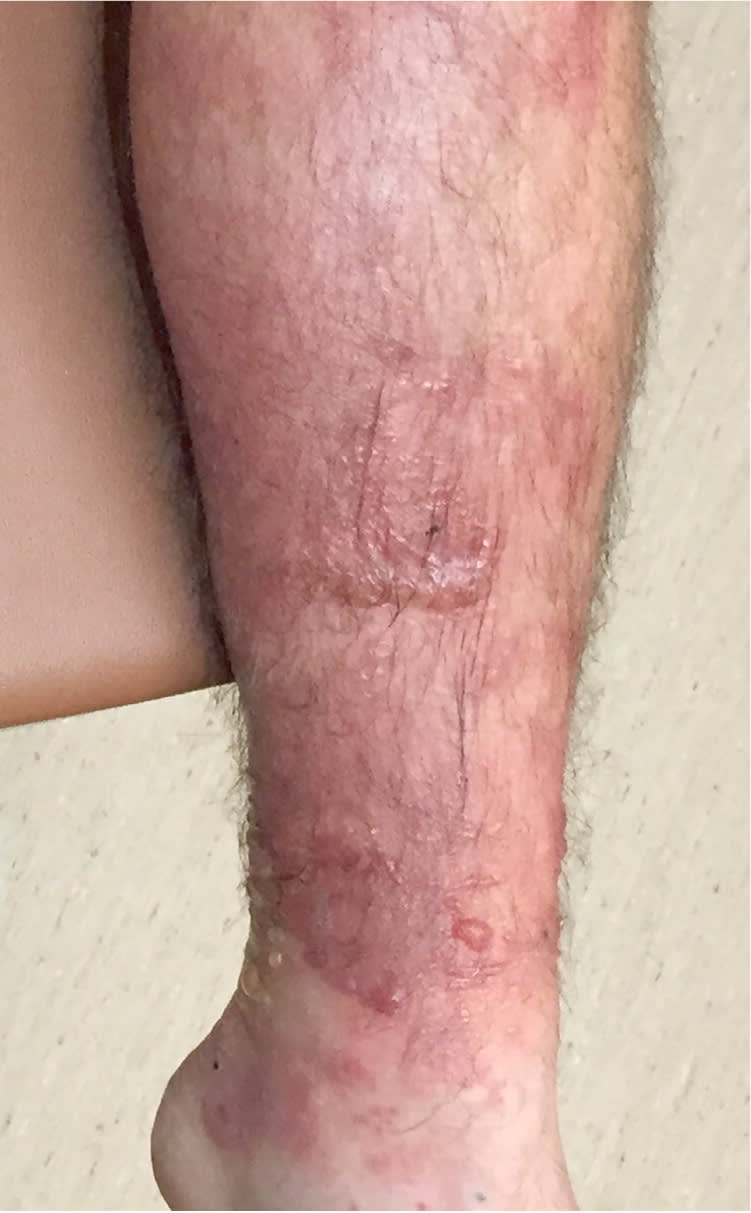
A 37-year-old man presented with a three-day history of itching and erythema on his right forearm and legs. There were bumps, blisters, and swelling in the area of the redness. He first noticed the rash on his forearm after walking back to his car from an overnight fishing trip. The next day, he noticed the rash on his legs. He was otherwise feeling well.
Physical examination revealed multiple papules and vesicles with erythema and edema (Figure 1). The erythema covered most of his legs, and some of the vesicles were oozing. There were no other physical findings. He was afebrile.
Figure 1.
Question
Based on the patient's history and physical examination findings, which one of the following is the most likely diagnosis?
A. Cellulitis.
B. Herpes simplex virus infection.
C. Poison ivy dermatitis.
D. Urticaria.
Discussion
The answer is C: poison ivy dermatitis, a type IV hypersensitivity allergic reaction to urushiol. Urushiol is the oleoresin contained in the stems, leaves, and roots of the poison ivy plant.1 In this case, the oleoresin first made contact with the right forearm and then with the legs shortly afterward.
Poison ivy is a type of Rhus dermatitis.1 The clinical presentation depends on the amount of the oleoresin that touches the skin, the way it touches the skin, and the patient's susceptibility and skin responsiveness. Presentation includes linear streaks that are pruritic, edematous, and erythematous, often with vesicles and large bullae on unclothed skin. Injury to the skin sometimes leaves a transient black mark, which is a sign of contact with the plant and the consequence of oxidized urushiol.2
The rash may occur eight hours to one or more weeks after exposure. The oleoresin is not found in the blister fluid and thus does not propagate the inflammation. Prevention entails washing the area with soap and water as soon as the exposure occurs, removing the surface oleoresin. The diagnosis of poison ivy dermatitis is clinical, with the intense, linear, vesicular outbreak being diagnostic.1 Treatment of the inflammation may include wet compresses and topical or oral steroids.1
Cellulitis is a bacterial infection affecting the skin and subcutaneous tissue.1 The condition presents as an enlarging red, warm, swollen, tender or painful plaque. The rash may affect a limited or extensive area.2 There is usually a history of trauma to the skin.
Herpes simplex virus infection may present as a primary or recurring outbreak. The infection presents as grouped vesicles on a red, swollen plaque that progress to pustules. The rash tends to recur near the initial site of infection. The rash is caused by type 1 or 2 herpesvirus infection.2
Urticaria, or hives, may present as an acute or chronic condition.2 The lesions are circumscribed, elevated, erythematous plaques, usually with central pallor. They may be round, oval, or serpiginous and range from 1 cm or less to several centimeters in diameter.3
Summary Table
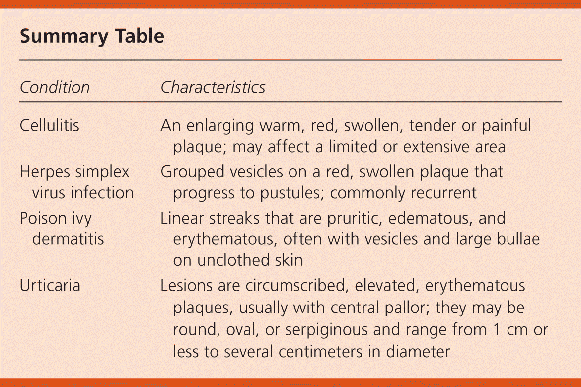
| Condition | Characteristics |
|---|---|
| Cellulitis | An enlarging warm, red, swollen, tender or painful plaque; may affect a limited or extensive area |
| Herpes simplex virus infection | Grouped vesicles on a red, swollen plaque that progress to pustules; commonly recurrent |
| Poison ivy dermatitis | Linear streaks that are pruritic, edematous, and erythematous, often with vesicles and large bullae on unclothed skin |
| Urticaria | Lesions are circumscribed, elevated, erythematous plaques, usually with central pallor; they may be round, oval, or serpiginous and range from 1 cm or less to several centimeters in diameter |
The opinions and assertions contained herein are the private views of the author and are not to be construed as official or as reflecting the views of the U.S. Air Force Medical Department or the U.S. Air Force at large.
