A 50-year-old woman presented with an asymptomatic white patch on her wrist that appeared two months earlier. There was no itching, pain, or swelling in the area. Her medical history included ulcerative colitis, asthma, carpal tunnel syndrome, and osteoarthritis. Her family history included a maternal aunt with extensive vitiligo.
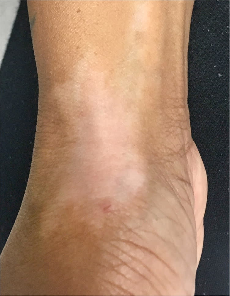
Physical examination revealed a single, irregularly shaped, well-defined, hypopigmented patch involving the lateral, dorsal, and ventral surfaces of the right wrist with an area of proximal linear extension along the ventral forearm (Figure 1). The area was smooth and not raised. The remainder of the physical examination was unremarkable.
Figure 1.
Question
Based on the patient's history and physical examination findings, which one of the following is the most likely diagnosis?
A. Idiopathic guttate hypomelanosis.
B. Pityriasis alba.
C. Steroid-induced hypopigmentation.
D. Vitiligo.
Discussion
The answer is C: steroid-induced hypopigmentation. A single patch of hypopigmented skin over a joint space leads to high suspicion for an exogenous condition. Hypopigmentation caused by corticosteroids presents as an irregularly shaped but well-defined hypopigmented patch. Further questioning of the patient revealed that she received a triamcinolone acetonide injection in the right carpometacarpal joint to treat osteoarthritis four months before presentation.
Triamcinolone acetonide is commonly used for intra-articular injections. It is a macromolecule with reduced solubility, which leads to more prolonged duration of action but a higher risk of hypopigmentation.1 Possible adverse effects of steroid injections include local irritation, infections, telangiectasia, cutaneous and subcutaneous atrophy, and epidermal dyspigmentation.1 Steroid-induced hypopigmentation is a common adverse effect of topical corticosteroids; however, it is less common following intra-articular steroid injections.2
Hypopigmentation usually occurs at the site of injection but may occur as linear rays, thought to be from lymphatic spread of suspended triamcinolone crystals.3 Most published cases of steroid-induced hypopigmentation occurred in black patients, although cutaneous hypopigmentation is less noticeable in patients with lighter skin tones and may be underreported.4 The reaction may be more cosmetically problematic in patients with darker skin types. Because hypopigmentation may occur months after an injection, the patient may not relate it to the steroid injection.4,5 Cutaneous hypopigmentation tends to resolve without treatment, although it may take several months to one year.4,5
Idiopathic guttate hypomelanosis is a common acquired benign skin condition in patients older than 50 years with dark skin tones. It typically presents as many small, scattered, well-demarcated, hypopigmented macules on sun-exposed areas of the extremities.6
Pityriasis alba is a common condition in children with a history of atopy. It presents as hypopigmented patches with fine scaling and ill-defined borders that typically affect the cheeks.7
Vitiligo is an acquired condition characterized by depigmented macules and patches due to loss of melanocytes. The lesions are usually symmetric and most often affect the face, neck, and scalp, or areas of repeated trauma.6
Summary Table
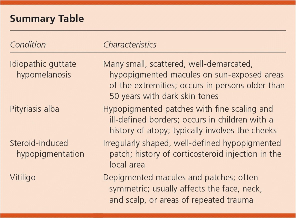
| Condition | Characteristics |
|---|---|
| Idiopathic guttate hypomelanosis | Many small, scattered, well-demarcated, hypopigmented macules on sun-exposed areas of the extremities; occurs in persons older than 50 years with dark skin tones |
| Pityriasis alba | Hypopigmented patches with fine scaling and ill-defined borders; occurs in children with a history of atopy; typically involves the cheeks |
| Steroid-induced hypopigmentation | Irregularly shaped, well-defined hypopigmented patch; history of corticosteroid injection in the local area |
| Vitiligo | Depigmented macules and patches; often symmetric; usually affects the face, neck, and scalp, or areas of repeated trauma |
