A 43-year-old man presented with a lesion on his right index finger. It had been present for a couple of years and was growing gradually. There was no history of trauma or infection to the area. He had no fever or weight loss.
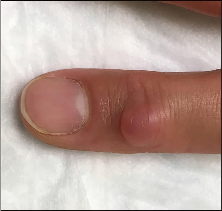
Physical examination revealed a soft, nontender, cystic lesion on the dorsum of the right second digit over the distal interphalangeal joint (Figures 1 and 2). There was no surrounding erythema or warmth, and the lesion was immobile with flexion and extension of the distal interphalangeal joint. There were positive findings on transillumination.
Figure 1.
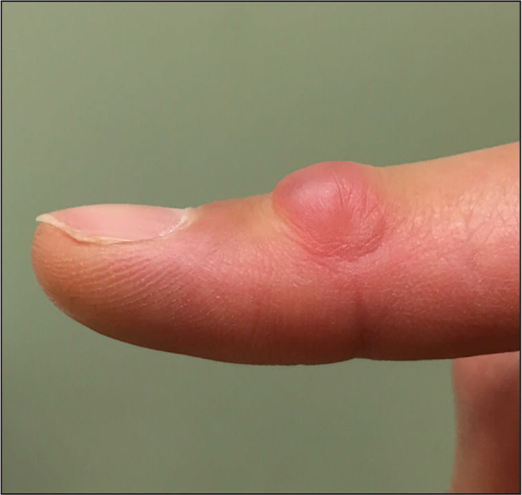
Figure 2.
Question
Based on the patient's history and physical examination findings, which one of the following is the most likely diagnosis?
A. Digital mucous cyst.
B. Fibrokeratoma.
C. Intradermal nevus.
D. Pyogenic granuloma.
Discussion
The answer is A: digital mucous cyst. Digital mucous cysts are solitary, flesh-colored lesions on the dorsal digits, usually between the distal interphalangeal joint and the proximal nail bed.1 They are benign ganglion cysts that are immobile with flexion and extension of the digits. They are more common on the fingers of the dominant hand but may also appear on toes.2 They can be a couple of millimeters to more than a centimeter in diameter. Digital mucous cysts usually present in middle-aged and older persons. Women are twice as likely as men to develop digital mucous cysts.1
There are two types of digital mucous cysts. One type develops from degenerative changes within the distal interphalangeal joint, and the other type is caused by fibroblast overproduction of hyaluronic acid.1 The two types cannot be differentiated clinically.3 Although the lesions are often asymptomatic, they may cause discomfort and decreased range of motion if located on or near a joint.1,3 When a cyst arises near the nail bed, it may cause tenderness and nail deformities, including damage to the nail matrix.1,3 Transillumination is helpful to determine if the lesion is a cyst.1 Digital mucous cysts may spontaneously regress, but most persist.1 Treatment ranges from observation, if the cyst is not affecting movement, to surgical removal.1,3
Fibrokeratomas are benign skin lesions, usually resulting from trauma. They often occur on the fingers, but may also appear on the hands or feet.4,5 The lesions consist of fibrous tissue and are often dome-shaped with a hyperkeratotic border.5
Intradermal nevi are often dome-shaped, although they can be verrucous or pedunculated.4 They range from dark brown to flesh-colored. The pigmentation in an individual lesion may change over time.4
Pyogenic granulomas are rapidly growing, acquired lesions of the skin and mucous membranes that are vascular in nature. They are often a result of trauma or hormonal changes. They are red and dome-shaped. They are most common in children, adolescents, and young adults.4,6
Selected Differential Diagnosis
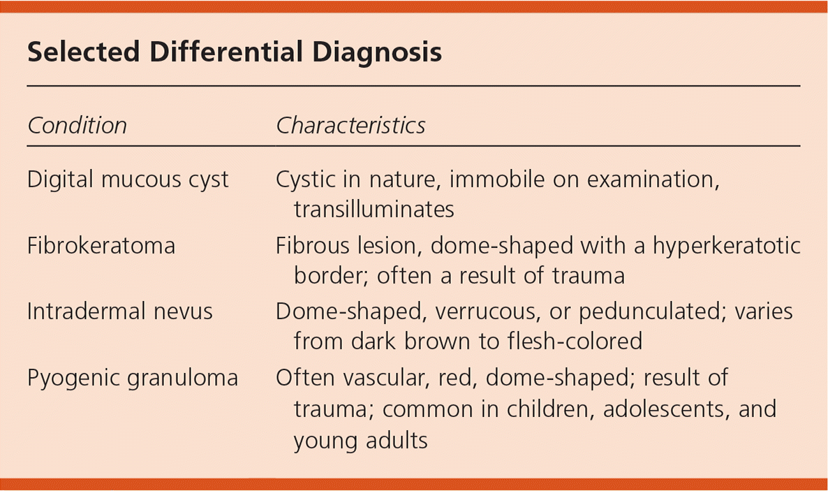
| Condition | Characteristics |
|---|---|
| Digital mucous cyst | Cystic in nature, immobile on examination, transilluminates |
| Fibrokeratoma | Fibrous lesion, dome-shaped with a hyperkeratotic border; often a result of trauma |
| Intradermal nevus | Dome-shaped, verrucous, or pedunculated; varies from dark brown to flesh-colored |
| Pyogenic granuloma | Often vascular, red, dome-shaped; result of trauma; common in children, adolescents, and young adults |
