A 45-year-old woman presented with a 10-day history of an erythematous, scaly rash on her back that developed after a period of intense sun exposure. She did not have a fever or chills, and there was no discharge from the site. The patient was otherwise healthy. She had a history of small, erythematous, scaly lesions on both elbows during adolescence that spontaneously resolved. Her family history was significant for maternal psoriasis.
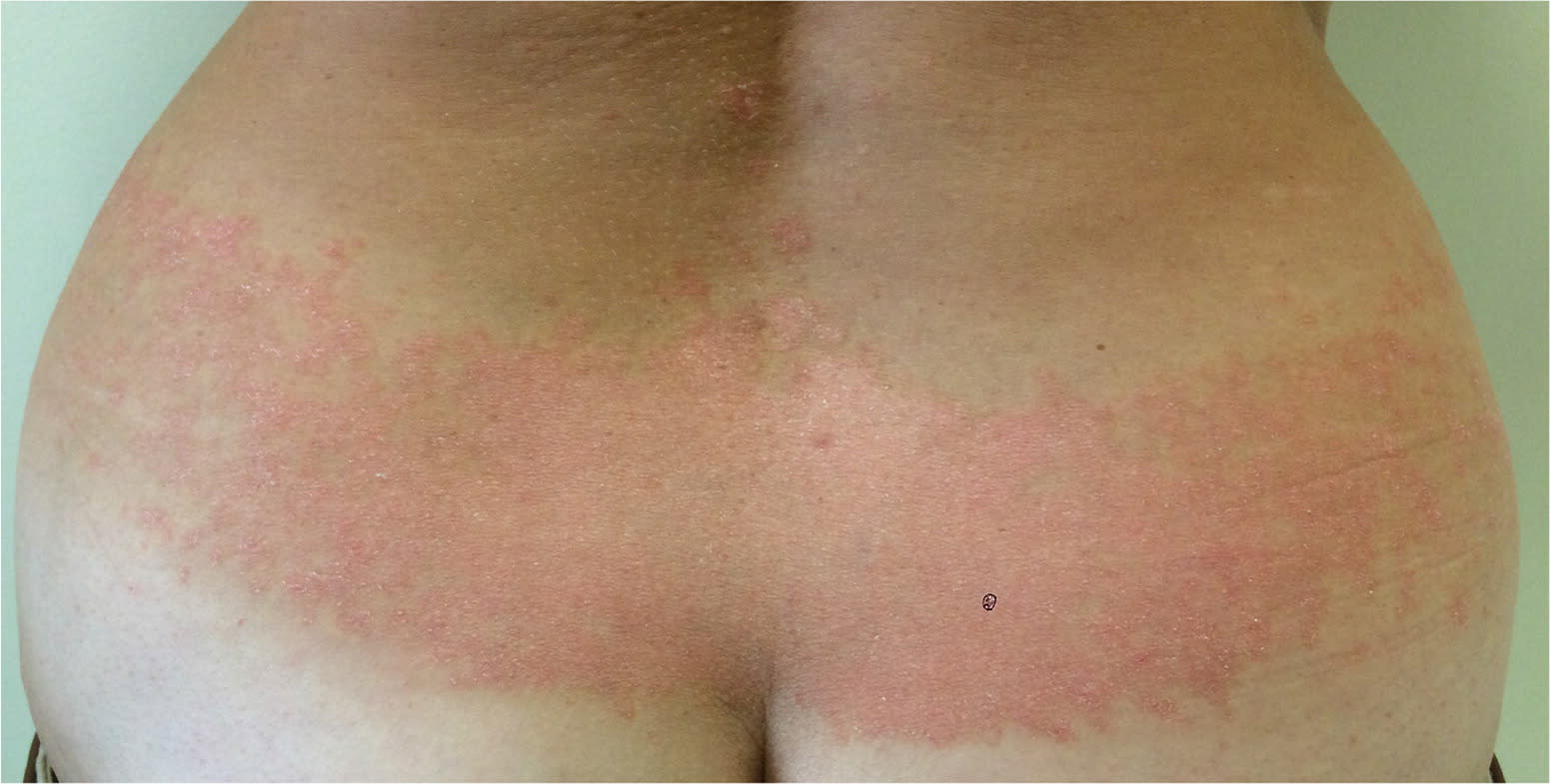
Physical examination revealed multiple erythematous, scaly plaques on the lower back just above the waistline (Figure 1). These lesions were in the area of sun exposure. There were no other lesions or physical findings.
Figure 1.
Question
Based on the patient's history and physical examination findings, which one of the following is the most likely diagnosis?
A. Herpes zoster.
B. Irritant contact dermatitis.
C. Koebner phenomenon.
D. Pityriasis rosea.
E. Tinea corporis.
Discussion
The answer is C: Koebner phenomenon. Koebner phenomenon is an isomorphous rash that is commonly associated with psoriasis and other skin diseases such as vitiligo and lichen planus.1 It occurs following local trauma in areas that are not involved with skin disease.2 The rash appears as clearly defined, erythematous, scaly plaques.3 Provoking factors can include physical trauma, friction, surgical incision, burns, and radiation exposure.4 Koebner phenomenon appears to be unrelated to the activity or severity of the associated skin disease.5
In general, the Koebner phenomenon rash appears 10 to 20 days after the local trauma.3 The reported incidence varies widely from 5% to 75%.5 No definitive pathogenic process has been identified.6 In this patient, the ultraviolet radiation from extensive sun exposure appears to be the trigger. Pathogenic immunologic processes may be enhanced by ultraviolet radiation. The patient's history of undiagnosed psoriasis may support this hypothesis.
Herpes zoster is a painful rash caused by reactivation of the varicella virus. It usually begins with a prodromal phase characterized by pain, itching, paresthesias, or dysesthesias in one dermatome. A few days later, a unilateral maculopapular rash appears on the affected area, which then evolves into clusters of small, tender vesicles and blisters in the corresponding dermatome.7,8
Irritant contact dermatitis is skin inflammation typically after exposure to an irritant. It presents as erythema, mild edema, and scaling in the pattern of the exposure. The rash is a nonspecific response to direct irritant damage that releases inflammatory mediators.7,8
Pityriasis rosea is a common skin disorder that occurs in otherwise healthy persons, most often children and young adults. The rash is a papulosquamous eruption that lasts six to eight weeks. It typically begins with a single salmon-colored herald patch on the trunk, followed by multiple lesions in a Christmas tree pattern. In some cases, it appears to occur after upper respiratory tract infection.7,8
Tinea corporis is a superficial dermatophyte infection characterized by an erythematous, scaly plaque with a raised, well-defined, advancing border. Following central resolution, the lesion appears annular in shape.7,8
Summary Table
| Condition | Characteristics of the rash |
|---|---|
| Herpes zoster | Clusters of small, tender vesicles and blisters |
| Irritant contact dermatitis | Erythema, mild edema, and scaling in the pattern of the irritant exposure |
| Koebner phenomenon | Clearly defined erythematous, scaly plaques following local trauma |
| Pityriasis rosea | Single salmon-colored herald patch on the trunk, followed by multiple lesions in a Christmas tree pattern |
| Tinea corporis | Annular, scaly plaque with a raised, well-defined, advancing border |
