Point-of-care ultrasonography (POCUS) refers to limited ultrasound protocols performed at the patient's bedside by a clinician to assess for many conditions such as aortic aneurysm and pleural effusion. The protocols usually answer a specific question (Table 1) that helps guide treatment and can be performed after a relatively brief training period. This is in distinction to consultative, comprehensive, or formal ultrasound examinations that are performed by sonographers and interpreted by radiologists with years of training in reading ultrasound results. POCUS, on the other hand, is a tool of the generalist. Its use was first introduced by emergency medicine physicians, but with technological advances allowing for smaller, pocket-sized ultrasound machines at lower costs, POCUS is becoming more accessible to family physicians. A feasibility study of the use of POCUS by family medicine residents and faculty who were given a handheld ultrasound machine and 16 total hours of training revealed users found it was easy to learn to use and improved diagnostic efficiency and accuracy, and patients were satisfied.1 In the study, 86% of participants agreed or strongly agreed that they would continue to use POCUS in their daily practices.1
TABLE 1. Questions Answered by Point-of-Care Ultrasonography That Are Applicable to Family Medicine
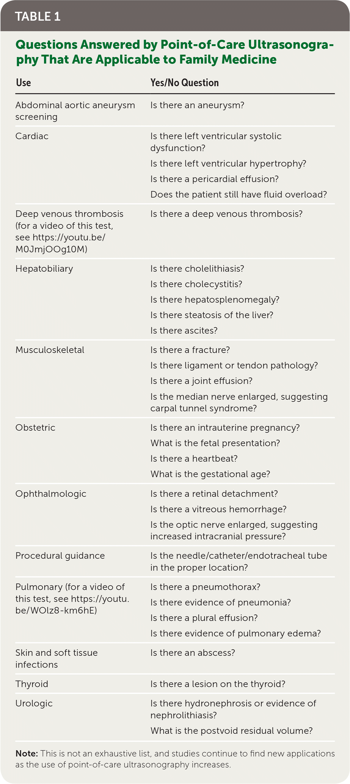
| Use | Yes/No Question |
|---|---|
| Abdominal aortic aneurysm screening | Is there an aneurysm? |
| Cardiac | Is there left ventricular systolic dysfunction? |
| Is there left ventricular hypertrophy? | |
| Is there a pericardial effusion? | |
| Does the patient still have fluid overload? | |
| Deep venous thrombosis (for a video of this test, see https://youtu.be/M0JmjOOg10M) | Is there a deep venous thrombosis? |
| Hepatobiliary | Is there cholelithiasis? |
| Is there cholecystitis? | |
| Is there hepatosplenomegaly? | |
| Is there steatosis of the liver? | |
| Is there ascites? | |
| Musculoskeletal | Is there a fracture? |
| Is there ligament or tendon pathology? | |
| Is there a joint effusion? | |
| Is the median nerve enlarged, suggesting carpal tunnel syndrome? | |
| Obstetric | Is there an intrauterine pregnancy? |
| What is the fetal presentation? | |
| Is there a heartbeat? | |
| What is the gestational age? | |
| Ophthalmologic | Is there a retinal detachment? |
| Is there a vitreous hemorrhage? | |
| Is the optic nerve enlarged, suggesting increased intracranial pressure? | |
| Procedural guidance | Is the needle/catheter/endotracheal tube in the proper location? |
| Pulmonary (for a video of this test, see https://youtu.be/WOlz8-km6hE) | Is there a pneumothorax? |
| Is there evidence of pneumonia? | |
| Is there a plural effusion? | |
| Is there evidence of pulmonary edema? | |
| Skin and soft tissue infections | Is there an abscess? |
| Thyroid | Is there a lesion on the thyroid? |
| Urologic | Is there hydronephrosis or evidence of nephrolithiasis? |
| What is the postvoid residual volume? | |
Note: This is not an exhaustive list, and studies continue to find new applications as the use of point-of-care ultrasonography increases.
There is mounting evidence that POCUS can help decrease the costs of care while improving patient access to care and safety. POCUS may reduce direct health care costs by serving as an initial triage tool to determine which patients may need more advanced imaging, thus decreasing the use of more expensive studies. A study comparing ultrasonography and computed tomography scans for evaluation of suspected nephrolithiasis in the emergency department found that initial testing with POCUS decreased the number of computed tomography scans by 59% without any change in outcomes.2 A 2008 study of Medicare data for musculoskeletal magnetic resonance imaging indicated that 45% of primary diagnoses could have been made with ultrasonography.3
Downstream costs from complications are a consideration as well. Ultrasonography has no radiation and no known direct adverse effects. Also, when used as an adjunct to help guide common procedures, such as venous access, thoracentesis, and arthrocentesis, ultrasonography has been shown to decrease rates of complications.4–6
In addition to being more cost-effective, POCUS is better than physical examination or plain radiography in many settings. When using POCUS, generalists are as accurate as cardiologists in assessing left ventricular systolic function, and even medical students are able to increase their diagnostic accuracy from 50% to 75%.7,8 POCUS is better than a physical examination in differentiating an abscess from cellulitis, changing management in 14% to 56% of cases.9 POCUS is superior to physical examination or chest radiography for making many lung diagnoses, including pleural effusion, pulmonary edema, pneumonia, and pneumothorax.10–15
Another benefit of POCUS in primary care is its ability to expedite and increase access to imaging. For example, abdominal aortic aneurysm screening using POCUS has a sensitivity of 99% to 100%, compared with screening in the radiology setting, and takes less than four minutes to complete.16,17 POCUS evaluation of the lower extremities for deep venous thrombosis has a sensitivity of 95% and specificity of 96%, compared with screening in the radiology setting, and can also be completed in less than four minutes.18,19
Given the benefits of different POCUS applications, interest in its use in the family physician's office is increasing. A 2014 survey of program directors found that only 2% of residency programs had a formal POCUS curriculum, but 29% indicated they had started one in the previous year and 11% were in the process of starting one.20 In 2016, the American Academy of Family Physicians (AAFP) Congress of Delegates passed a resolution encouraging all family medicine residency programs to include POCUS as part of their training and for the AAFP to increase continuing medical education offerings that incorporate POCUS training.21 The AAFP has since created a curriculum guideline for POCUS in graduate medical education.22 Resources for additional information on POCUS education and implementation are included in Table 2.
TABLE 2. Resources for Point-of-Care Ultrasonography Education and Implementation
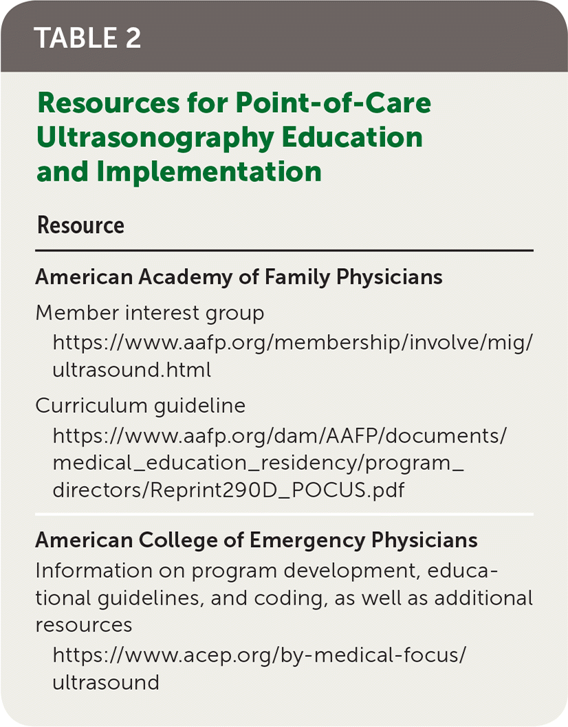
| Resource | |
|---|---|
| American Academy of Family Physicians | |
| Member interest group | |
| Curriculum guideline | |
| American College of Emergency Physicians | |
| Information on program development, educational guidelines, and coding, as well as additional resources |
