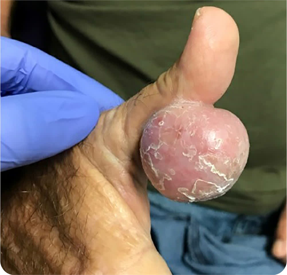
A 65-year-old man presented with an erythematous mass on his thumb that had been slowly enlarging over 10 years. The mass was nontender but bled occasionally and increasingly interfered with daily activities. The patient did not have fever, chills, or night sweats.
Physical examination revealed a large, semifirm, mobile mass on the volar aspect of the right thumb (Figure 1). The lesion was not ulcerated. There was no lymphadenopathy. The affected hand had normal strength, sensation, and range of motion.
FIGURE 1
Plain radiography showed a soft tissue mass without evidence of a foreign body. Magnetic resonance imaging showed an exophytic superficial soft tissue mass measuring 3.7 × 3.0 × 3.7 cm without evidence of internal fat, cystic nature, foreign body, or involvement of the underlying tendon. The mass was excised, and pathologic examination showed diffuse subcutaneous fibrosis and granulomatous inflammation comprised of epithelioid histiocytes.
Question
Based on the patient’s history, physical examination findings, and additional testing, which one of the following is the most likely diagnosis?
- A. Actinomycetoma.
- B. Dermatofibroma.
- C. Foreign body granuloma.
- D. Giant cell tumor of the tendon sheath.
- E. Invasive squamous cell carcinoma.
Discussion
The answer is A: actinomycetoma. Actinomycetoma is a painless, chronic, inflammatory, granulomatous, suppurative cutaneous lesion caused by Actinomadura, Nocardia, or Streptomyces bacteria, which are commonly found in soil. Mycetomas can be differentiated into actinomycetomas (caused by non–acid-fast bacterial infections) and eumycetoma (caused by fungal infections).1
Mycetomas present clinically as nodules, abscesses, fistulae, and swelling. The lesions usually involve the lower extremities but can also occur on the hands. Tendons and nerves are spared until late in the disease process. The disease is typically spread via lymphatic and hematologic systems. Mycetomas can cause distortions, disfigurement, and disability.2 If left untreated, they can cause superinfections, osteomyelitis, and ankylosis.1,2 Rarely, mycetomas are aggressive and can progress systemically, involving pelvic organs, the spinal cord, and lungs, with a high mortality rate.2
Gram stain shows thin filaments (0.5 to 1 μm) with actinomycetomas,3 but thicker fungal hyphae are seen with eumycetomas. Classic histologic findings are suppurative granulomas with neutrophilic infiltrates surrounded by palisaded histiocytes.1 Methenamine silver stain is used to highlight filamentous bacteria.
Although evidence varies, common treatments for actinomycetomas include dapsone, streptomycin, and trimethoprim/sulfamethoxazole. The treatment duration and dosage are dependent on disease severity and tissue involvement.1,3 Surgical debulking may be an option for larger, more burdensome masses but has an increased risk of recurrence.1
Dermatofibromas are benign fibrous histiocytomas of the skin, usually related to trauma in the affected area. Lesions are typically firm and less than 1 cm in diameter. Histology shows proliferation of spindle-shaped fibrous cells and histiocytoid cells in a storiform pattern.4
Foreign body granulomas are caused by penetrating objects, typically made of glass, metal, or wood. Radiographic visualization depends on the material’s radiodensity. Magnetic resonance imaging is the best modality for identification of nonmetallic sources. Histology shows granulation tissue with fibroblasts and inflammatory infiltrates of lymphocytes, plasma cells, histiocytes, and giant cells.5
Giant cell tumor of the tendon sheath is the second most common benign neoplasm of the hand. It is a painless lesion of the soft tissues. Histology shows these lesions are composed of multinucleated giant cells, histiocytes, fibrotic material, and hemosiderin deposits.6
Invasive squamous cell carcinomas appear ulcerated and can be patchy, papulonodular, papillomatous, or exophytic lesions.7 They can also resemble their precursor, actinic keratosis lesions, but with evidence of infiltration of cells into the dermis. Histologically, they have atypical nuclei, pleomorphism, keratinization, or sometimes a pearl-like dermal nest. Patients may also present with weight loss, night sweats, and pain.8
SUMMARY TABLE
| Condition | Characteristics |
|---|---|
| Actinomycetoma | Painless, chronic, inflammatory, granulomatous, suppurative cutaneous lesion caused by Actinomadura, Nocardia, or Streptomyces bacteria; histology shows granulomas with neutrophilic infiltrate surrounded by palisaded histiocytes; Gram stain commonly shows thin filaments |
| Dermatofibroma | Benign fibrous histiocytomas of the skin; histology shows proliferation of spindle-shaped fibrous cells and histiocytoid cells in a storiform pattern |
| Foreign body granuloma | Granuloma formation caused by a penetrating object, such as glass, metal, or wood; object often visible on plain radiography or magnetic resonance imaging; histology shows granulation tissue with fibroblasts and inflammatory infiltrates |
| Giant cell tumor of the tendon sheath | Painless lesion of the soft tissues; histology shows these lesions are composed of multinucleated giant cells, histiocytes, fibrotic material, and hemosiderin deposits |
| Invasive squamous cell carcinoma | Ulcerated; can be patchy, papulonodular, papillomatous, or exophytic; histology shows atypical nuclei, pleomorphism, keratinization, or sometimes a pearl-like dermal nest; may be associated with weight loss, night sweats, and pain |
