

| Test | Indication | Population | Cost* |
|---|---|---|---|
| Temporal artery ultrasonography | New onset of headache | Adults 50 years and older | $861 |
*—Cost according to the Centers for Medicare and Medicaid Services.12
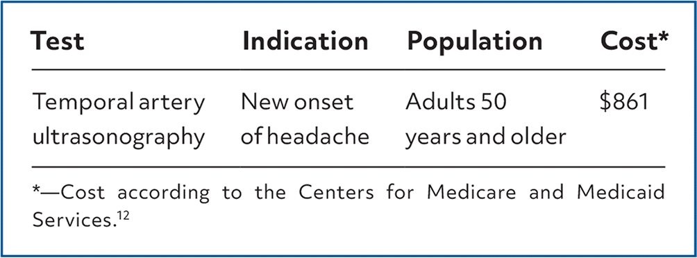
Giant cell arteritis (GCA) should be suspected in patients 50 years and older with new onset of headaches. Erythrocyte sedimentation rate (ESR) and C-reactive protein (CRP) levels are often elevated in patients with GCA.1 Temporal artery biopsy and vascular ultrasonography are two modalities that can be used to diagnose GCA. Ultrasonography of the temporal artery is the preferred method of diagnosis in Europe. Key findings that suggest GCA on ultrasonography include the halo sign (hypoechoic arterial wall thickening due to edema), the compression sign (inability of the technician to compress the lumen of the superficial temporal artery due to vessel wall inflammation), vessel stenosis, or total occlusion.2 There is no standard cutoff for halo sign thickness that is universally considered diagnostic of GCA.3
ACCURACY
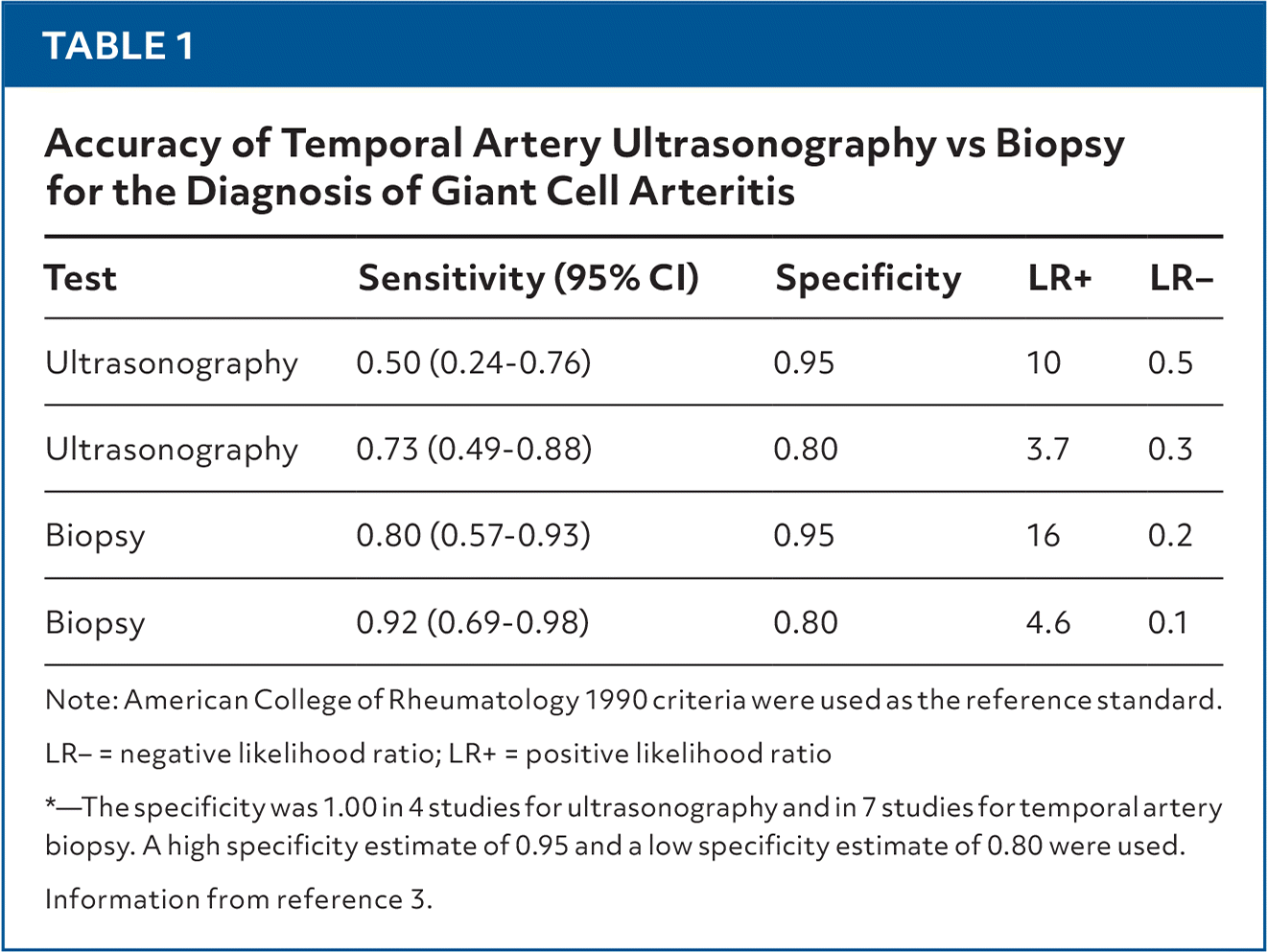
Reported sensitivities and specificities for both unilateral temporal artery biopsy and ultrasonography in the diagnosis of GCA vary widely. To investigate whether ultrasonography could be used as triage for performing biopsy, a 2024 systematic review evaluated the accuracy of the halo sign on ultrasonography using the 1990 American College of Rheumatology criteria as the reference standard (positive for at least three of the following: 50 years of age or older at disease onset, new headache, temporal artery abnormality, elevated ESR, abnormal findings on temporal artery biopsy).3 Temporal artery ultrasonography was investigated in 15 studies including 1,479 patients. Of these patients, 617 (41.7%) had GCA. The evidence regarding sensitivity (median = 0.78) and specificity (median = 0.91) was of very low certainty due to bias, imprecision, and inconsistency in halo thickness threshold among studies. Temporal artery biopsy was compared with ultrasonography in 11 studies including 808 patients; 460 patients (56.9%) had GCA (Table 1).3 The evidence on the sensitivity of ultrasonography compared with biopsy was very low due to overestimation of specificity. Specificity was one of the criteria used in the reference standard, leading to incorporation bias.
TABLE 1. Accuracy of Temporal Artery Ultrasonography vs Biopsy for the Diagnosis of Giant Cell Arteritis
| Test | Sensitivity (95% CI) | Specificity | LR+ | LR− |
|---|---|---|---|---|
| Ultrasonography | 0.50 (0.24–0.76) | 0.95 | 10 | 0.5 |
| Ultrasonography | 0.73 (0.49–0.88) | 0.80 | 3.7 | 0.3 |
| Biopsy | 0.80 (0.57–0.93) | 0.95 | 16 | 0.2 |
| Biopsy | 0.92 (0.69–0.98) | 0.80 | 4.6 | 0.1 |
Note: American College of Rheumatology 1990 criteria were used as the reference standard.
LR− = negative likelihood ratio; LR+ = positive likelihood ratio
*—The specificity was 1.00 in 4 studies for ultrasonography and in 7 studies for temporal artery biopsy. A high specificity estimate of 0.95 and a low specificity estimate of 0.80 were used.
Information from reference 3.
A 2016 multicenter cohort study in Europe compared temporal artery ultrasonography and biopsy in 381 patients with suspected GCA. Patients underwent ultrasonography and biopsy within 10 days of starting treatment for suspected GCA. Performance of the two tests were evaluated against a reference diagnosis determined by the clinician. Ultrasonography had a sensitivity of 54% (95% CI, 0.48–0.60), and biopsy had a sensitivity of 39% (95% CI, 0.33–0.46).4 Biopsy had a specificity of 100% (95% CI, 0.97–1.0), and ultrasonography had a specificity of 81% (95% CI, 0.73–0.88).4 Incorporation bias was also a limitation in this study. Four studies used the same halo cutoff of greater than 0.3 mm. Excluding one outlier (sensitivity of 0.10, specificity of 1.0), the other three studies had similar accuracy, with sensitivity ranging from 73% to 88% and specificity ranging from 97% to 100%.
Read the full article
Get immediate access, anytime, anywhere.
Choose a single article, issue, or full-access subscription.
Earn up to 14 CME credits per issue.
