

A 30-year-old man presented with a left shoulder injury from weight lifting 3 days prior. He was bench-pressing 125-lb dumbbells when his left hand drifted wide, and he felt his shoulder dislocate anteriorly. He was able to immediately reduce his left shoulder by pushing it posteriorly with his right hand. The patient’s shoulder then gradually lost mobility, and the pain worsened to 10 out of 10. He then needed to support his left arm. Radiography results were negative.
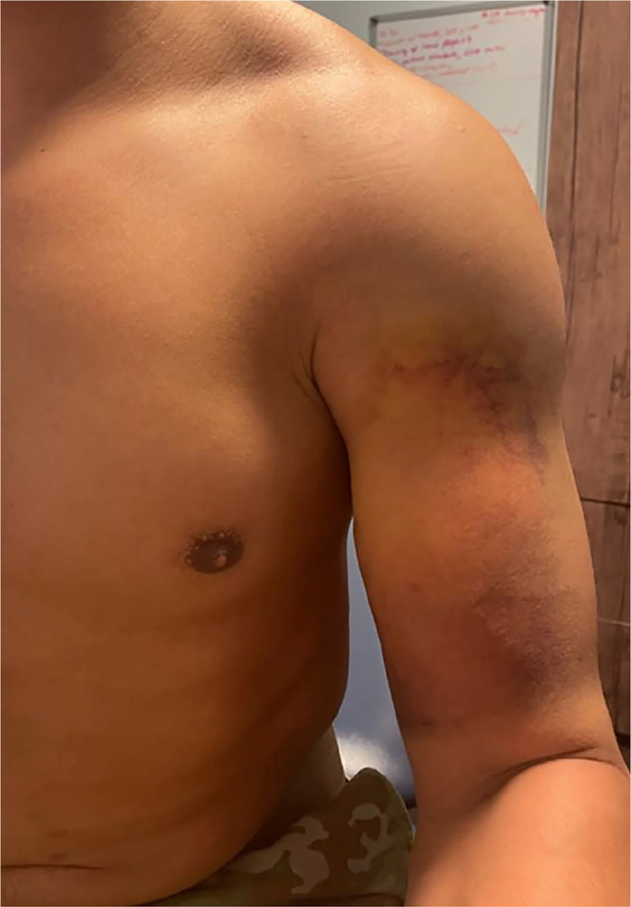
Physical examination showed significant ecchymosis overlying his left biceps (Figure 1). Pain limited the physical examination, particularly flexion and abduction of the left shoulder. The patient was not able to tolerate manual muscle testing of his left shoulder; however, strength was grossly 80% in all directions. The patient’s elbow flexion was unaffected and did not exacerbate shoulder pain.
FIGURE 1
QUESTION
Based on the patient’s history and physical examination, which one of the following is the most likely diagnosis?
- A. Acromioclavicular joint sprain (shoulder separation).
- B. Complete tear of the proximal biceps brachii.
- C. Pectoralis major tendon rupture.
- D. Shoulder instability.
DISCUSSION
The answer is C: pectoralis major tendon rupture. Traumatic tears to the pectoralis major tendon or myotendinous junction feature swelling and ecchymosis over the anterolateral chest wall and proximal arm. Typical causes of injury include heavy bench pressing and hyperabduction.1 Shoulder injuries account for one-third of musculoskeletal presentations to primary care, and the differential diagnosis of acute shoulder pain should include pectoralis injury.2 Physical examination reveals weakness with shoulder flexion and internal rotation and loss of normal muscle contour or anterior axillary fold, which is best observed with the arm abducted 90 degrees. Asymmetry of the chest wall is noted in the prayer position, with the patient pressing their hands together to highlight isometric contraction of the muscle.3
Magnetic resonance imaging is the preferred imaging modality, with a sensitivity for pectoral tears ranging from 51% to 100% and specificity from 64% to 75%, depending on the type of tear.4,5 Musculoskeletal ultrasonography can be used when magnetic resonance imaging is unavailable, but its accuracy is unclear.6,7 Surgical management within 6 weeks of injury reduces the risk of postoperative complications and maximizes outcomes.8
Acromioclavicular joint sprain (shoulder separation) is usually traumatic, such as from a direct blow to the lateral shoulder. Visual abnormalities are present only with grade 2 or 3 sprains (ligament tears). Physical examination findings include tenderness on direct palpation. Other tests include the cross arm test and the scarf test.
Complete tear of the proximal biceps brachii is associated with visual abnormality of the biceps (Popeye deformity), weakness or pain with resisted elbow flexion, and tenderness on palpation around the insertions of the biceps.
Shoulder instability is a clinical diagnosis describing compromise of structures supporting the humeral head, such as the structures and dynamic muscular stabilizers (eg, rotator cuff muscles). Physical examination findings include the sulcus sign and positive apprehension-relocation test result.
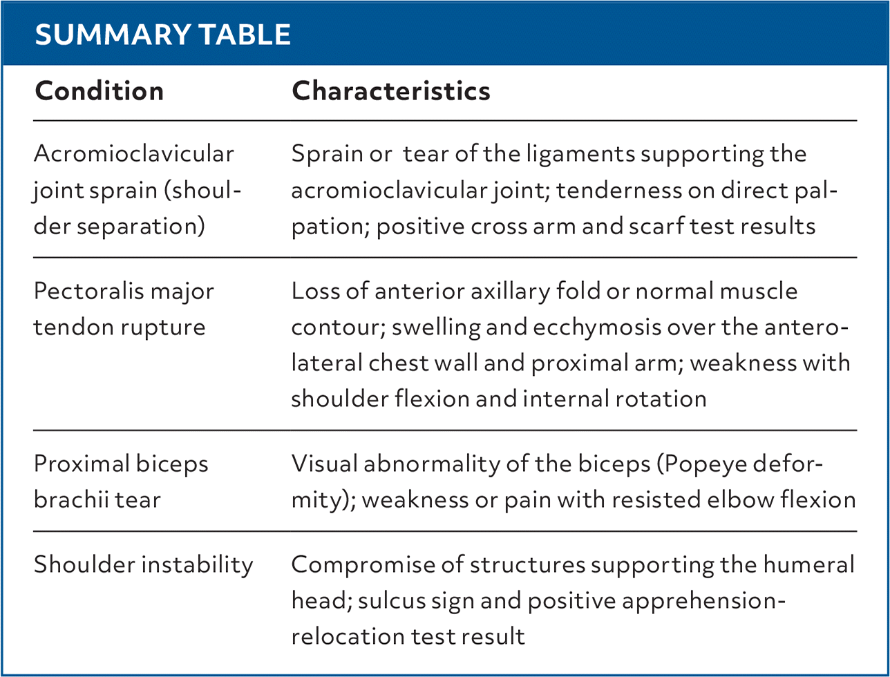
SUMMARY TABLE
| Condition | Characteristics |
|---|---|
| Acromioclavicular joint sprain (shoulder separation) | Sprain or tear of the ligaments supporting the acromioclavicular joint; tenderness on direct palpation; positive cross arm and scarf test results |
| Pectoralis major tendon rupture | Loss of anterior axillary fold or normal muscle contour; swelling and ecchymosis over the anterolateral chest wall and proximal arm; weakness with shoulder flexion and internal rotation |
| Proximal biceps brachii tear | Visual abnormality of the biceps (Popeye deformity); weakness or pain with resisted elbow flexion |
| Shoulder instability | Compromise of structures supporting the humeral head; sulcus sign and positive apprehension-relocation test result |
Read the full article
Get immediate access, anytime, anywhere.
Choose a single article, issue, or full-access subscription.
Earn up to 14 CME credits per issue.
