

A 59-year-old man presented with a brown-black lesion on the hard palate. The lesion was associated with pain and caused difficulty eating. The lesion started as a small, black-pigmented area 2 years ago and grew gradually. The patient was a 10-pack-year smoker.
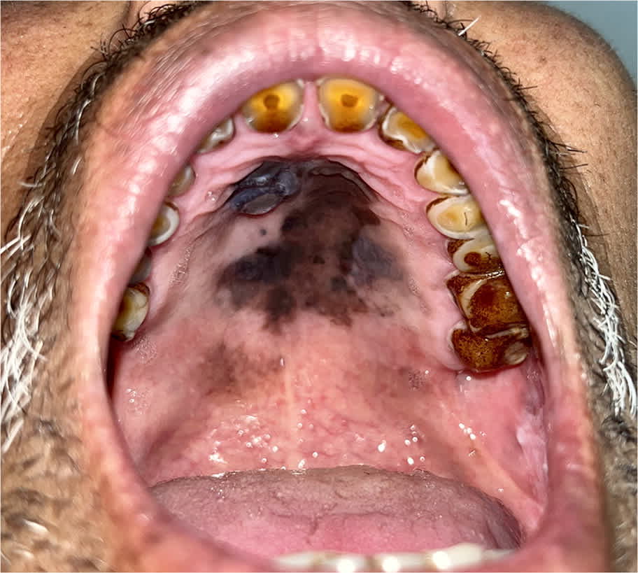
Examination of the oral cavity revealed poor oral hygiene and nicotine staining of the teeth. A sharply demarcated, variably pigmented lesion with superimposed nodularity and irregular borders covered a large area of the hard palate (Figure 1). The patient’s upper cervical lymph nodes were bilaterally enlarged (2 cm in diameter) and were mobile, firm, and nontender.
FIGURE 1
QUESTION
Based on the patient’s history and physical examination, which one of the following is the most likely diagnosis?
- A. Drug-induced melanosis.
- B. Melanocytic nevus.
- C. Melanotic macule.
- D. Palatal melanoma.
- E. Smoker’s melanosis.
DISCUSSION
The answer is D: palatal melanoma. Oral melanoma usually presents with three key features: a brown-black pigmented plaque, light-brown macular area, and central area with nodularity. Smoking has an unclear relationship with the development of head and neck mucosal melanoma, but it is correlated with increased melanocyte proliferation in the oral mucosa, increasing the risk of pigmented oral lesions.1
Melanoma of the oral cavity comprises 1% of all melanomas, with a higher incidence in men older than 50.2 The most common sites are the maxillary gingiva and hard palate. Pigment variations may include melanotic and amelanotic areas within the same lesion. Other common symptoms include ulceration, pain, and paresis. Early detection through clinical evaluation is crucial for optimal management and improved outcomes.
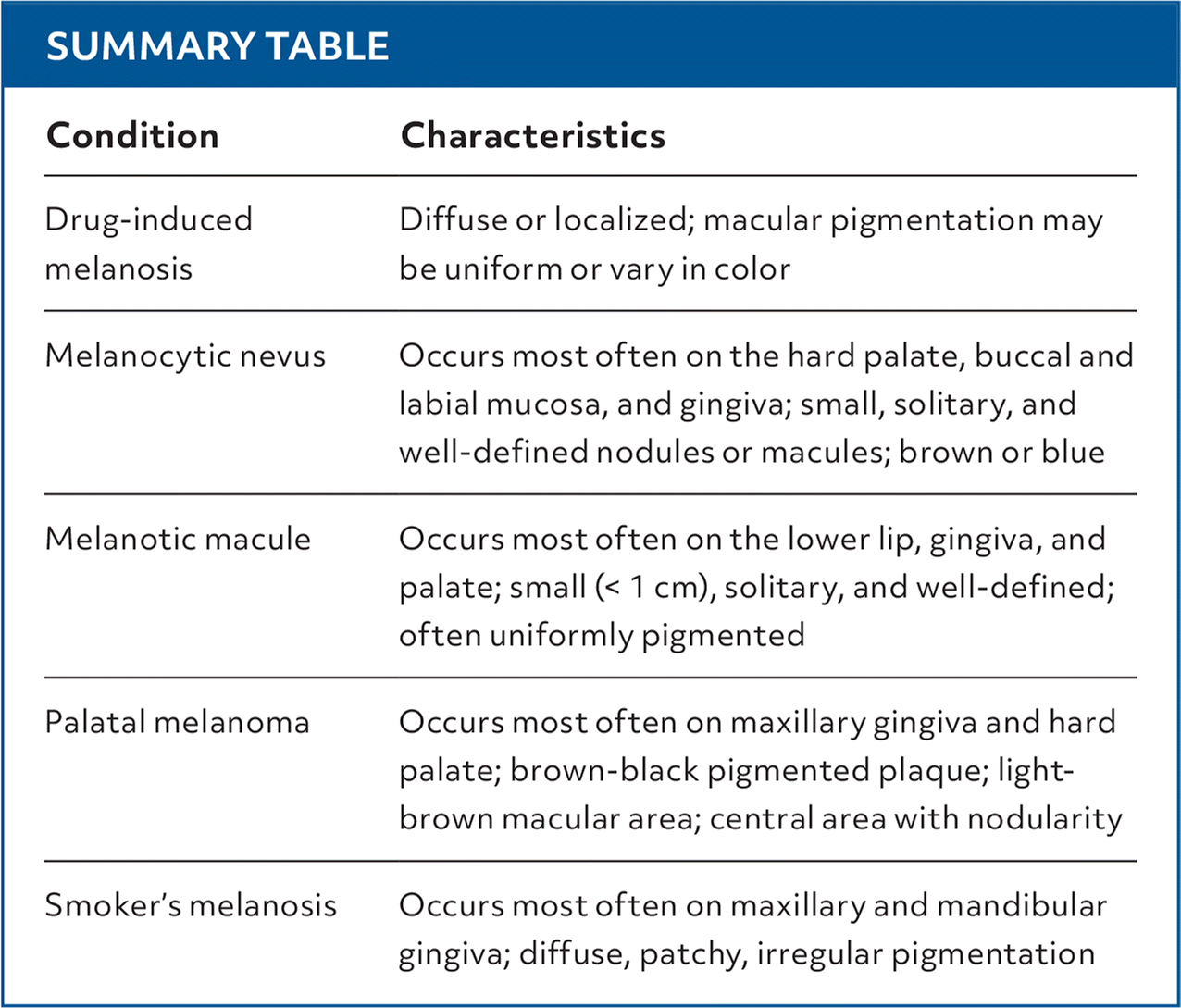
Drug-induced melanosis can be diffuse or localized. The macular pigmentation may be uniform or vary in color. Some drugs cause a specific pattern of pigmentation, such as hydroxychloroquine triggering palatal mucosal pigmentation.3
Oral melanocytic nevi commonly develop in individuals who are at least 30 years of age. Typically asymptomatic, these lesions may manifest as small, solitary, and well-defined nodules or macules that are brown or blue.4 The lesions can affect any mucosal site but are most common on the hard palate, buccal and labial mucosa, and gingiva.
Melanotic macules are the most common oral mucosal lesions that originate from melanocytes. They are small, solitary, well-defined, and often uniformly pigmented. They tend to be more prevalent in adult women. Melanotic macules can occur on any mucosal site, most often the lower lip, gingiva, and palate.5 Melanotic macules rarely exceed 1 cm in diameter.
Smoking is linked to abnormal pigmentation of the oral mucosa (melanosis) due to nicotine staining and poor oral hygiene. Smoker’s melanosis presents as diffuse, patchy, irregular pigmentation that primarily affects the maxillary and mandibular gingiva. Melanosis is not considered a precancerous condition.6
SUMMARY TABLE
| Condition | Characteristics |
|---|---|
| Drug-induced melanosis | Diffuse or localized; macular pigmentation may be uniform or vary in color |
| Melanocytic nevus | Occurs most often on the hard palate, buccal and labial mucosa, and gingiva; small, solitary, and well-defined nodules or macules; brown or blue |
| Melanotic macule | Occurs most often on the lower lip, gingiva, and palate; small (< 1 cm), solitary, and well-defined; often uniformly pigmented |
| Palatal melanoma | Occurs most often on maxillary gingiva and hard palate; brown-black pigmented plaque; light-brown macular area; central area with nodularity |
| Smoker’s melanosis | Occurs most often on maxillary and mandibular gingiva; diffuse, patchy, irregular pigmentation |
Read the full article
Get immediate access, anytime, anywhere.
Choose a single article, issue, or full-access subscription.
Earn up to 14 CME credits per issue.
