

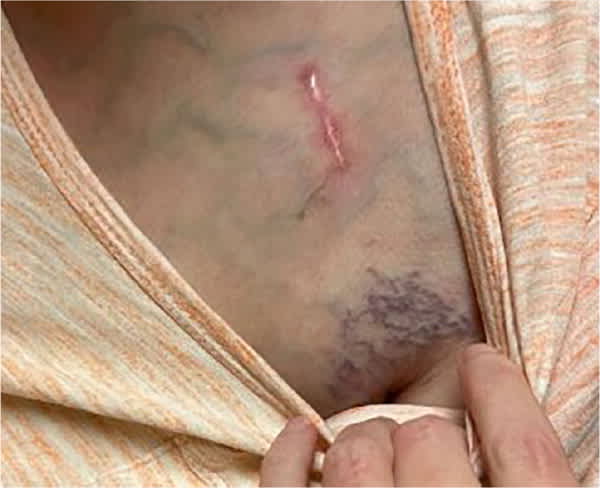
A 66-year-old woman presented 2 months after implantation of a biventricular permanent pacemaker. She had no immediate complications after the procedure. Four weeks after the procedure, she noted new varicose veins in the upper portion of her left arm and pectoral area, followed by swelling that started in her proximal upper arm and spread distally. The swelling was not reduced by arm elevation. The patient did not have pain, numbness, or weakness in the arm; swelling in other areas; increased shortness of breath; or change in systemic symptoms.
Physical examination revealed a well-healing pacemaker scar site, prominent varicose veins, and notable swelling of her left upper extremity (Figure 1 and Figure 2).
FIGURE 1
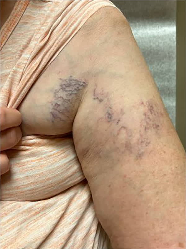
FIGURE 2
QUESTION
Based on the patient's history and physical examination, which one of the following is the most likely diagnosis?
- A. Deep vein thrombosis.
- B. Lead-related venous obstruction.
- C. Lymphedema.
- D. Superior vena cava syndrome.
DISCUSSION
The answer is B: lead-related venous obstruction. Venous occlusion secondary to pacemaker lead placement is a relatively common complication; it occurs when there is venous narrowing of 50% or more.1 Patients can be asymptomatic, have limb swelling and venous distension on the side of the device, or, in severe cases, develop superior vena cava syndrome.2 The most significant risk factor is the presence of multiple leads.3
When venous obstruction is suspected, venous Doppler ultrasonography is recommended to evaluate for acute thrombosis. Contrast venography is considered the diagnostic standard. Most cases are managed conservatively, but some patients may require lead extraction or percutaneous revascularization.2
Deep vein thrombosis is less common in the upper extremities than in the lower extremities, but the incidence is increasing due to more frequent use of central venous catheters and pacemakers. An estimated 6% of acute thromboses involve upper extremities.4 The condition typically presents with extremity pain and swelling, and diagnosis is made with venous ultrasonography.5 Deep vein thrombosis following pacemaker implantation typically occurs within 6 months of the procedure and is treated with anticoagulation.6
Lymphedema is caused by accumulation of protein-rich fluid in interstitial spaces. Upper extremity lymphedema most commonly results from breast cancer treatment. The condition is diagnosed clinically and presents in a spectrum of severity from edema that resolves with limb elevation to significant nonpitting edema with acanthosis and skin thickening. Treatment ranges from complex decongestive therapy to operative intervention.7
Superior vena cava syndrome is caused by compression of the superior vena cava in the anterior or middle mediastinum. The resulting increased pressure causes dilation and development of distended neck and chest veins, facial edema, plethora, cough, hoarseness, and headache. Malignancy is the most common cause. Superior vena cava syndrome associated with intravascular devices is rare, presenting in 0.1% to 0.3% of patients with obstruction.8 Management of superior vena cava syndrome is difficult, but percutaneous stenting is the most common approach.9
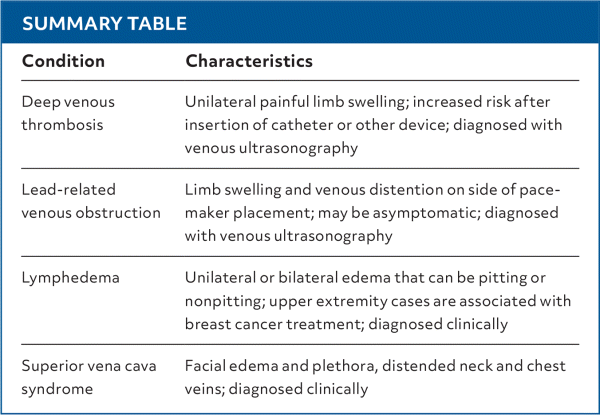
SUMMARY TABLE
| Condition | Characteristics |
|---|---|
| Deep venous thrombosis | Unilateral painful limb swelling; increased risk after insertion of catheter or other device; diagnosed with venous ultrasonography |
| Lead-related venous obstruction | Limb swelling and venous distention on side of pacemaker placement; may be asymptomatic; diagnosed with venous ultrasonography |
| Lymphedema | Unilateral or bilateral edema that can be pitting or nonpitting; upper extremity cases are associated with breast cancer treatment; diagnosed clinically |
| Superior vena cava syndrome | Facial edema and plethora, distended neck and chest veins; diagnosed clinically |
Read the full article
Get immediate access, anytime, anywhere.
Choose a single article, issue, or full-access subscription.
Earn up to 14 CME credits per issue.
