

CLINICAL QUESTION
For women with dense breasts, which supplemental study (magnetic resonance imaging [MRI], automated whole breast ultrasonography, or contrast-enhanced mammography) had the greatest yield for cancer following a normal mammogram result?
BOTTOM LINE
Contrast-enhanced mammography and MRI have high rates of cancer detection (1.6%–1.7%) in women with heterogeneously or extremely dense breasts, even after a normal mammogram result. Contrast-enhanced mammography may be a less expensive option for large-scale screening programs, although it comes with a small risk of contrast reaction. Further follow-up from this study and the previous Dutch study is necessary to understand the screening impact on interval cancers, overdiagnosis, and mortality. (Level of Evidence = 1b)
SYNOPSIS
Women with increased breast density are at higher risk of having an invasive breast cancer being detected when it is larger. Ultrasonography and MRI have been proposed as supplemental screening for these women, although it is not formally endorsed by any organization. Breast density is graded on a 4-point scale, and the UK researchers identified 9,361 women with grade C (heterogeneously dense) or grade D (uniformly dense) breast density who had a normal screening mammogram. The women were randomized to one of four groups: no supplemental screening (usual care), MRI, automated whole breast ultrasonography, or contrast-enhanced mammography. The latter uses a low-ionic iodinated intravenous contrast media. The median age of participants was 56 years, approximately 80% had grade C density, 20% had grade D density, and more than 90% completed the assigned supplemental screening within 6 months. Cancer detection and recall rates were not available for the usual care group, but that group will contribute data to subsequent publications that will report the rates of interval cancers and cancers at round two of screening. Results of the first round of screening are summarized in Table 1.
TABLE 1. Comparison of Breast Cancer Imaging Modalities
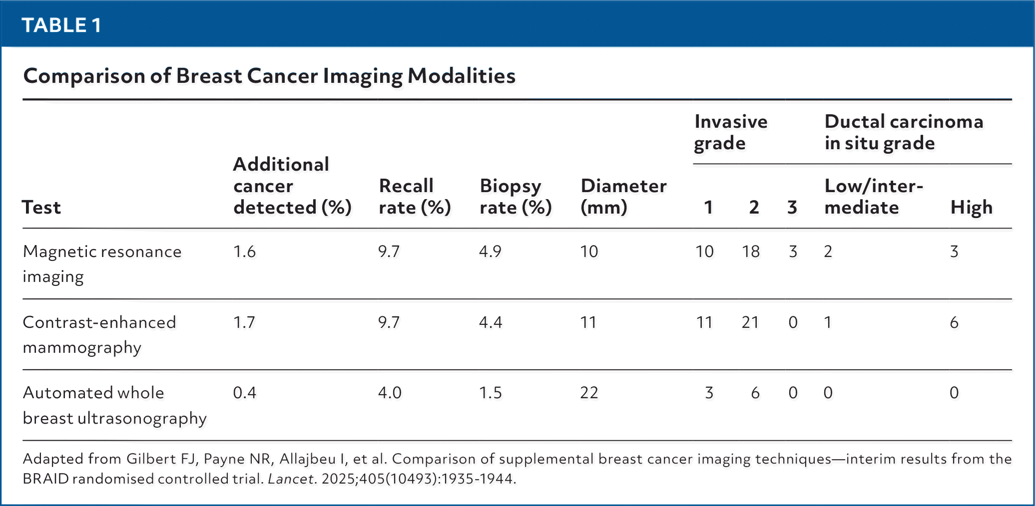
| Test | Additional cancer detected (%) | Recall rate (%) | Biopsy rate (%) | Diameter (mm) | Invasive grade | Ductal carcinoma in situ grade | |||
|---|---|---|---|---|---|---|---|---|---|
| 1 | 2 | 3 | Low/intermediate | High | |||||
| Magnetic resonance imaging | 1.6 | 9.7 | 4.9 | 10 | 10 | 18 | 3 | 2 | 3 |
| Contrast-enhanced mammography | 1.7 | 9.7 | 4.4 | 11 | 11 | 21 | 0 | 1 | 6 |
| Automated whole breast ultrasonography | 0.4 | 4.0 | 1.5 | 22 | 3 | 6 | 0 | 0 | 0 |
Adapted from Gilbert FJ, Payne NR, Allajbeu I, et al. Comparison of supplemental breast cancer imaging techniques—interim results from the BRAID randomised controlled trial. Lancet. 2025;405(10493):1935–1944.
MRI and contrast-enhanced mammography had much higher rates of cancer detection and smaller tumor diameter than automated whole breast ultrasonography, although these were accompanied by higher rates of recall and biopsy. The rate of cancer detection for MRI was identical to that reported by a similar Dutch study that also found lower rates of interval cancer, although the Dutch study only included women with grade D density. Most of the invasive cancers were low or intermediate grade, whereas most of the ductal carcinomas in situ were high grade and required treatment. In addition to high rates of recall and biopsy, there were one severe, six moderate, and 17 minor contrast media reactions among 2,235 women in the contrast-enhanced mammography group.
Read the full article
Get immediate access, anytime, anywhere.
Choose a single article, issue, or full-access subscription.
Earn up to 14 CME credits per issue.
