

A 62-year-old man presented as a new patient. He had no symptoms except a slowly growing left abdominal mass. It started developing approximately 8 years earlier, but he had not sought previous medical treatment. The patient did not have chest pain, shortness of breath, or bowel or bladder symptoms. His medical history was significant for hypertension and coronary artery disease. He had no history of abdominal surgery.
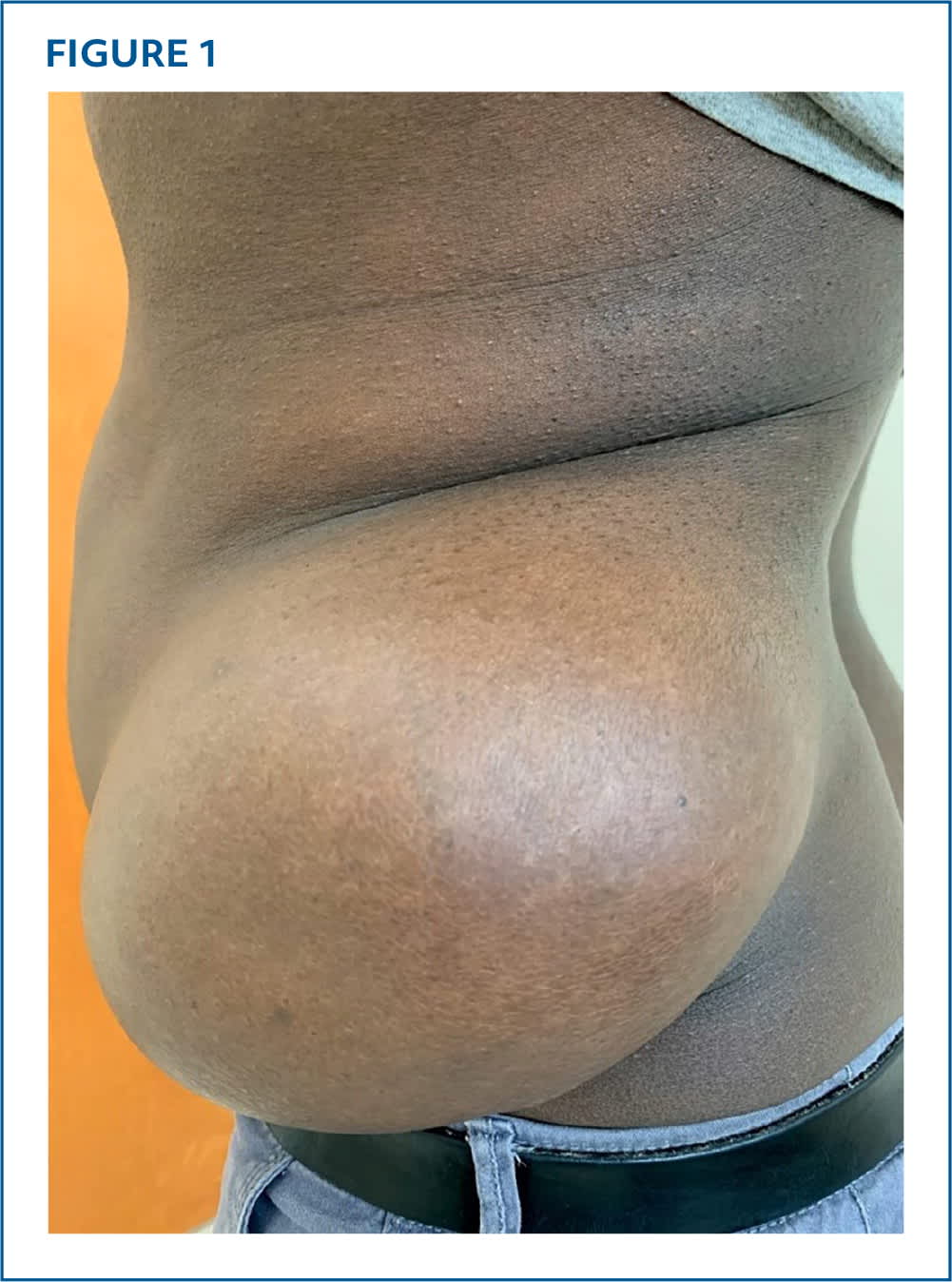
Physical examination revealed a large soft mass protruding from his left flank that was nontender, mobile, and subcutaneous (Figure 1). Urinalysis, complete metabolic profile, and complete blood count results were normal.
FIGURE 1
QUESTION
Based on the patient's history and physical examination, which one of the following is the most likely diagnosis?
- A. Angiolipoma.
- B. Epidermoid cyst.
- C. Lipoma.
- D. Liposarcoma.
- E. Rhabdomyosarcoma.
DISCUSSION
The answer is C: lipoma. The patient was counseled that he most likely had a noncancerous fatty tumor. Ultrasonography confirmed the presence of a solid subcutaneous mass. The patient noted that his brother had previously been treated for a similar mass. Surgery was performed, and a 17.5 × 13.5 × 11 cm mass weighing 1,259 g was removed. Histological examination demonstrated encapsulated benign adipose tissue with several focal areas of fat necrosis.
Lipomas are the most common benign soft tissue neoplasms and may have a genetic component. Most often, they occur in subcutaneous tissue of the trunk, neck, forearms, and proximal extremities. They present as a mobile, nontender mass with the characteristic clinical finding of the slippage sign (ie, as the examiner's fingers slide off the edge of the mass, the mass is felt to slip away). This sign indicates the mass is freely mobile and not tethered to the surrounding tissue. Ultrasonography can help rule out an epidermoid cyst, but definitive diagnosis requires surgical biopsy. Because progression to liposarcoma is rare, surgical removal is necessary only for cosmetic purposes.1
Angiolipomas are benign tumors that typically occur in adolescents and young adults. Their usual location is on the forearm or chest wall, and they present as multiple slow-growing, well-circumscribed masses. Angiolipomas are typically tender, which can help differentiate them from lipomas. A defining feature of angiolipomas is histological findings of adipose cells with a vascular component.1
Epidermoid cysts are the most common skin cysts. They are believed to form from disruption in the normal sebaceous environment and can contain components of sebum, such as comedones. They are located in areas with substantial hair follicles and can be caused by epidermal trauma. Fine-needle aspiration with cytology can confirm the diagnosis.2
Liposarcomas are common soft tissue sarcomas. They develop in adipose tissue and are usually found in the retroperitoneum and extremities. These tumors are progressive and can result in abdominal pain from compression. They can grow very large and may exceed 20 cm in diameter.3
Rhabdomyosarcomas are believed to be tumors of striated muscle precursor cells. They commonly present in children and young adolescents as rapidly progressive, high-grade malignancies. Physical examination reveals a nontender mass with overlying erythema that can occur anywhere on the body.4
Read the full article
Get immediate access, anytime, anywhere.
Choose a single article, issue, or full-access subscription.
Earn up to 14 CME credits per issue.
