

An 11-year-old boy presented with a rash on his back, abdomen, arms, and legs that had been present for several years. The rash was nonpruritic, but the patient experienced occasional irritation. He was previously treated with ketoconazole cream without improvement. Treatment with topical hydrocortisone and moisturizer was also ineffective. The patient's mother reported he was otherwise healthy and growing well.
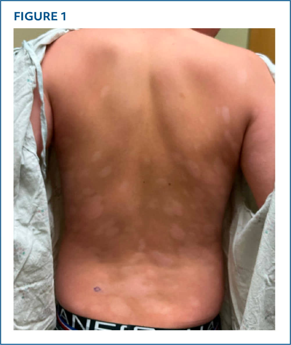
Physical examination revealed well-demarcated hypopigmented macules and patches that were coalescing on his back, abdomen, buttocks, medial upper arms, and posterior thighs (Figure 1). Some of the lesions had mild erythema and over-lying scale, and a few were subtly atrophic. A hypopigmented lesion on his right posterior thigh showed erythema with over-lying scale and follicular prominence. Wood lamp evaluation showed hypopigmentation. Biopsies of two representative lesions were obtained.
FIGURE 1
QUESTION
Based on the patient's history and physical examination, which one of the following is the most likely diagnosis?
- A. Atopic dermatitis.
- B. Hypopigmented mycosis fungoides.
- C. Tinea versicolor.
- D. Vitiligo.
DISCUSSION
The answer is B: hypopigmented mycosis fungoides, a cutaneous T-cell lymphoma. This is the most common variant of mycosis fungoides in children. It typically presents with persistent round to irregular, hypopigmented, nonatrophic patches with overlying fine scale. Follicular lesions may also be present. The patches usually appear on the trunk, buttocks, and limbs. They are generally asymptomatic but may be mildly pruritic.1 A biopsy is required for diagnosis, and multiple biopsies from different lesions may be necessary.
The histopathology of hypopigmented mycosis fungoides is similar to that of classic mycosis fungoides, including epidermotropism of lymphocytes, perivascular and periadnexal infiltrate, haloed lymphocytes, patchy lichenoid infiltrate, psoriasiform epidermal hyperplasia, and dermal melanophages.2 Additionally, although both conditions express CD2, CD3, CD4, and CD5, classic mycosis fungoides is CD8 negative and hypopigmented mycosis fungoides is CD8 positive. Polymerase chain reaction testing can detect T-cell gene rearrangements to further confirm diagnosis.2
Cutaneous lesions are often the only manifestation of hypopigmented mycosis fungoides, and treatments directed at the skin are typically effective with a favorable prognosis.2 Treatments include phototherapy, immunotherapy, ultraviolet radiation therapy, and chemotherapy. Topical treatments such as corticosteroids, retinoids, and histone deacetylase inhibitors are also effective.3
Atopic dermatitis is an inflammatory skin condition affecting up to 20% of children.4 Lesions are typically erythematous with intensely pruritic, scaly papules and papulovesicles. In children, lesions can be widespread but most often involve the face, trunk, flexor surfaces, and volar aspects of the wrists and ankles. Postinflammatory hyperpigmentation and hypopigmentation are common.4 Diagnosis is made clinically, and biopsy is generally not recommended. Atopic dermatitis can be differentiated from hypopigmented mycosis fungoides by the presence of intense pruritus.
Tinea versicolor is a cutaneous fungal infection caused by the yeast Malassezia furfur. It presents as hyperpigmented, hypopigmented, or erythematous macules and patches most commonly on the trunk, upper extremities, neck, and face. The lesions are often asymptomatic but can be mildly pruritic. Diagnosis is made with a potassium hydroxide preparation demonstrating short hyphae and yeast cells in a “spaghetti and meatball” pattern.5
Vitiligo is an autoimmune condition that causes the loss of melanocytes. Lesions are asymptomatic, nonscaly, depigmented macules or patches. They can occur anywhere on the body but are most common in the periorificial areas, lips, distal extremities, groin, and areas of friction. Wood lamp evaluation shows lesions that appear bright blue or with white fluorescence demonstrating focal depigmentation. Dermoscopy can also show perifollicular pigmentation and telangiectasias.6 Biopsy is not necessary for diagnosis.
Read the full article
Get immediate access, anytime, anywhere.
Choose a single article, issue, or full-access subscription.
Earn up to 14 CME credits per issue.
