

A 59-year-old postmenopausal woman presented for a routine gynecological examination. She reported mild dyspareunia but did not have dysuria, hematuria, or pelvic or abdominal pain. She had experienced no uterine bleeding since completing menopause 4 years earlier. Her last Papanicolaou test result 4 years earlier was negative for human papillomavirus but showed atypical squamous cells of undetermined significance.
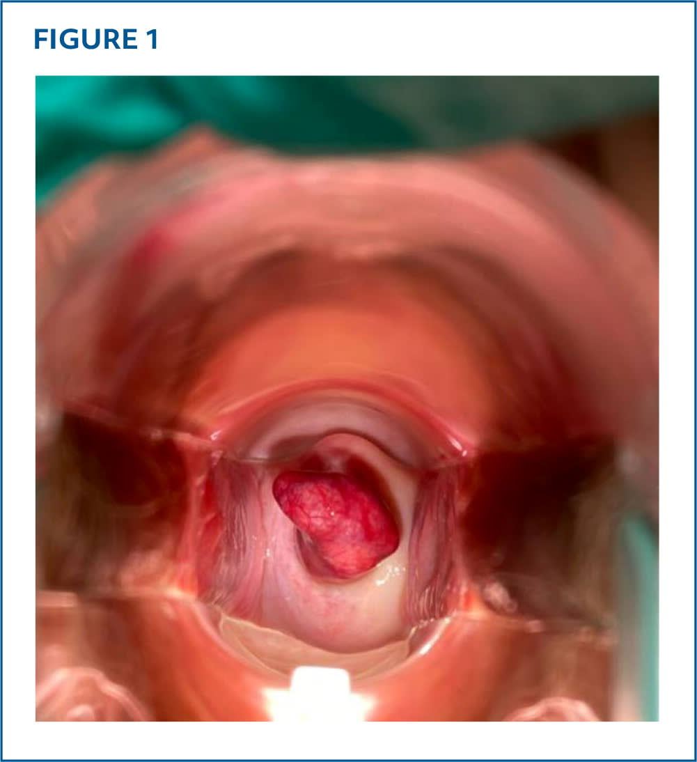
A speculum examination revealed a fleshy mass 3 cm in diameter protruding from the cervical os (Figure 1). No vaginal discharge or bleeding was noted.
FIGURE 1
QUESTION
Based on the patient's history and physical examination, which one of the following is the most likely diagnosis?
- A. Cervical carcinoma.
- B. Cervical polyp.
- C. Endometrial carcinoma.
- D. Endometrial polyp.
- E. Prolapsing submucosal fibroid (leiomyoma).
DISCUSSION
Answer is D: endometrial polyp. Endometrial polyps are localized proliferations of the endometrial glands and stroma that develop within the uterine cavity. They can range in size from a few millimeters to several centimeters. They may appear as a solitary growth or as multiple lesions that occupy the entire endometrial space. Although endometrial polyps can occur at any age, they are most common between 40 and 49 years. Risk of malignant transformation has been reported to be as high as 13%.1
Transvaginal ultrasonography showed a probable endometrial polyp. The patient then underwent saline infusion sonohysterography and hysteroscopy, which verified the findings. Hysteroscopic excision demonstrated a complex polyp originating from the left lateral uterine fundus. Pathological examination confirmed a benign endometrial polyp with no hyperplasia.
The exact etiology of endometrial polyps is unknown; however, they may be associated with endometrial hyperplasia mediated by unopposed estrogen. Other risk factors include use of tamoxifen, obesity, postmenopausal status, and hormone replacement therapy. Genetic syndromes that increase cancer risk are also associated with polyp development. Patients may be asymptomatic but commonly present with uterine bleeding or intermenstrual spotting. Other associated symptoms include abdominal pain, pelvic pain, and infertility.
Transvaginal ultrasonography is the initial diagnostic imaging study. Findings may appear nonspecific with diffuse or localized echogenic endometrial thickening. Increased endometrial thickness of more than 4 mm is related to endometrial pathology, including polyps.2 Tissue sampling should be considered due to the risk of malignancy.
Asymptomatic, low-risk endometrial polyps can often be managed conservatively with observation because they may resolve spontaneously, especially in premenopausal women. Periodic monitoring via ultrasonography is recommended. The optimal indications and timing for polyp removal in asymptomatic patients is uncertain. Larger polyps (more than 1.5 cm) are less likely to regress and may warrant removal, particularly when symptoms are present. Hysteroscopic polypectomy is the preferred treatment, allowing for both removal and histologic evaluation, especially because polyps can coexist with endometrial cancer. Hysterectomy is a more definitive option, but it is invasive and should be considered only after thorough counseling due to its risks and costs.
A cervical carcinoma appears as an irregular, ulcerated, friable mass on the cervix with disruption of cervical stroma. Risk factors include sexually transmitted infections (especially with human papillomavirus), genetic propensity, and tobacco use. Associated symptoms include postcoital bleeding and vaginal discharge. Risk of malignancy is high. A normal Pap test result with the absence of significant dysplasia makes this diagnosis less likely.3
Read the full article
Get immediate access, anytime, anywhere.
Choose a single article, issue, or full-access subscription.
Earn up to 14 CME credits per issue.
