

A 49-year-old man presented for his annual physical examination and had concerns about a nail abnormality. His medical history was significant for chronic pain disorder managed with oxycodone, and he was a 20-pack-year smoker. He ambulated with the assistance of a cane. There was no history of trauma to the nail. The abnormality had developed in the past 3 months.
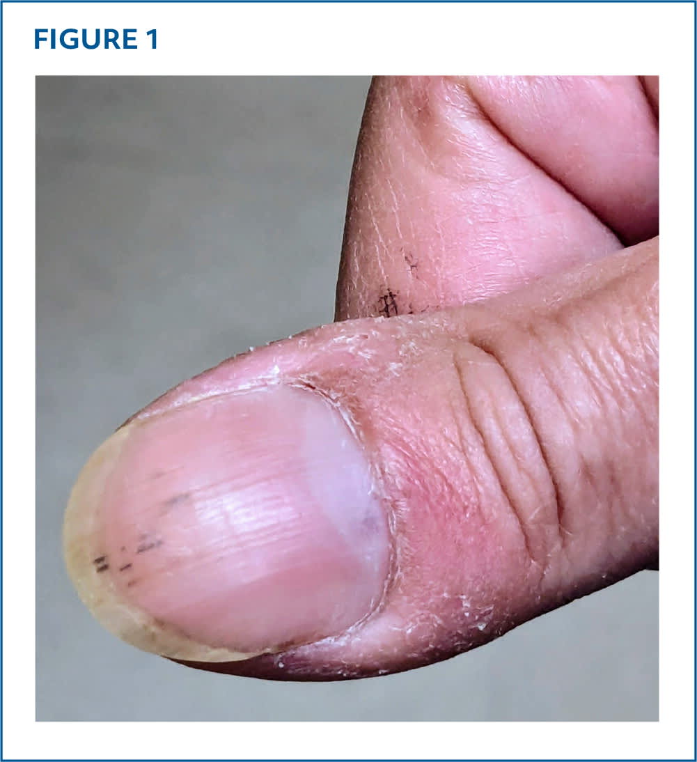
Physical examination revealed clubbing of all fingernails and thin, hyperpigmented, nonblanchable, vertical streaks in the distal half of the right thumbnail (Figure 1). The patient had a regular heart rate and rhythm with no murmurs, rubs, or gallops, and his lungs were bilaterally clear to auscultation. He was afebrile.
FIGURE 1
QUESTION
Based on the patient's history and physical examination, which one of the following is the most likely diagnosis?
- A. Melanonychia striata.
- B. Splinter hemorrhage.
- C. Subungual hematoma.
- D. Subungual melanoma.
DISCUSSION
The answer is B: splinter hemorrhage. Splinter hemorrhages present as nonblanchable, thin, linear, reddish-brown to black longitudinal streaks within the nail bed. The streaks typically do not span the entire nail bed. They may occur on one or more nails. Cases that are limited to a single nail typically have benign causes, most often trauma. Some cases are idiopathic. The prevalence of splinter hemorrhages is increased in manual workers, individuals who wear acrylic fingernails, and those who use a cane.1
When splinter hemorrhages present on multiple nails or with other symptoms, they may be associated with conditions such as infectious endocarditis, dermatoses, connective tissue disease, vasculitis, or kidney failure.1 Certain medications, including tyrosine kinase inhibitors, nitrofurantoin, and bevacizumab, have also been associated with splinter hemorrhages.1–3
Melanonychia striata presents as a longitudinal streak or irregular band of nonblanchable, brown, black, or tan discolorations that span the entire nail bed. In most cases, it is benign and associated with darker skin tone or increased melanin production due to pregnancy, medications, or infections. Less commonly, it is caused by melanocyte hyperplasia that may result from melanocytic nevus or invasive subungual melanoma. Due to the broad differential diagnosis of melanonychia striata, any malignant etiology should be ruled out.4,5
Subungual hematoma presents as irregular black or brown discoloration under the nail matrix caused by pooling of blood. This can occur following traumatic injury to the nail bed, commonly direct blows or crushing injuries. As blood accumulates, it causes increased pressure and pain underneath the nail. Trephination, a procedure where a small hole is drilled in the nail bed to relieve pressure, is recommended for drainage of acute hematomas within 48 hours of onset.6
Subungual melanoma is a subtype of acral-lentiginous melanoma. It appears as a nonblanchable, longitudinal streak or irregular brown-black discoloration spanning the entire nail bed. The big toe and thumb are affected in 75% to 90% of cases. Features include bands greater than 3 mm, irregular borders, and progressive changes in size or pigmentation. Periungual pigmentation of the proximal or lateral nail folds may be present and is an important diagnostic clue. Definitive diagnosis is made by full-thickness biopsy of the nail bed.7
Read the full article
Get immediate access, anytime, anywhere.
Choose a single article, issue, or full-access subscription.
Earn up to 14 CME credits per issue.
