

A 74-year-old woman presented with changes in the color of her nails and skin that started 15 years earlier. She had a history of type 2 diabetes, obstructive sleep apnea, recent lower extremity deep venous thrombosis treated with apixaban (Eliquis), and endometrial cancer. She consumed a daily colloidal silver supplement (6 oz) for viral illnesses and minor skin infections. She lived on a farm and used well water.
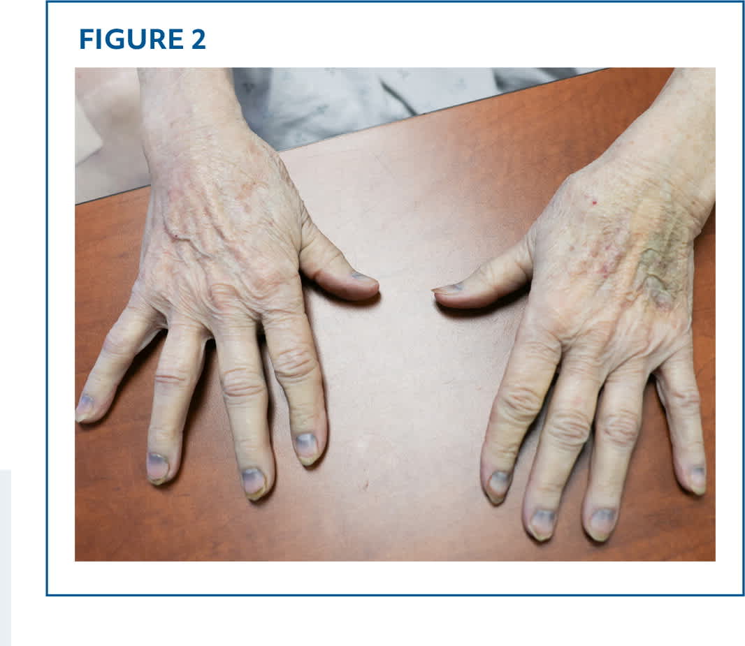
Physical examination revealed blue-gray discoloration of her nail lunulae and skin (Figure 1 and Figure 2). There was no evidence of cyanosis. Iron studies and measurement of arterial blood gases, ceruloplasmin, and urinary copper levels had normal results.
FIGURE 1
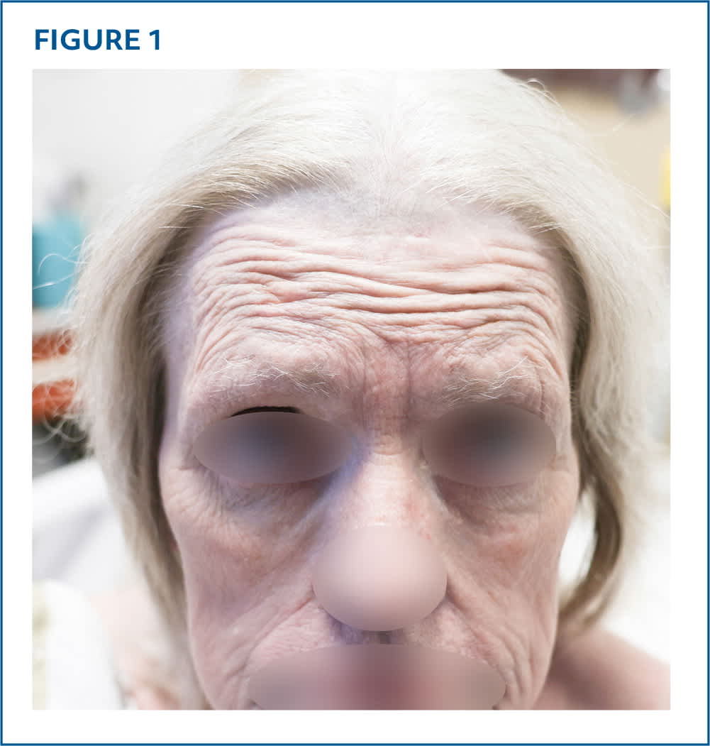
FIGURE 2
QUESTION
Based on the patient’s history and physical examination, which one of the following is the most likely diagnosis?
A. Alkaptonuria.
B. Argyria.
C. Hemochromatosis.
D. Methemoglobinemia.
E. Wilson disease.
DISCUSSION
The answer is B: argyria, discoloration of tissues and organs caused by chronic absorption of products that contain silver. Silver deposition typically occurs in the skin and can manifest as localized dark-blue macules to a generalized gray-blue skin tone. The discoloration is most prominent in sun-exposed areas.1 Localized cutaneous argyria can occur when silver makes direct contact with an area of skin on a regular basis, as seen in occupational exposures. This is most common in silversmiths or welders but may also result from repeated or prolonged contact with acupuncture needles, jewelry, or topical medications such as silver nitrate.2 When silver intake exceeds the excretory capabilities of the liver and kidneys, the excess metal is deposited in the tissues and can lead to generalized argyria. This can occur in virtually every organ, including the skin.3
Despite the lack of evidence of effectiveness, silver is sometimes believed to be a cure-all in alternative and homeopathic medicine.4
Argyria is a benign condition, although it can be cosmetically distressing. Treatment options are limited because the skin changes are often permanent. Treatment with hydroquinone, oral penicillamine, or dermabrasion is not effective. Success has been reported with laser techniques.5
Alkaptonuria is a rare genetic disorder in which an enzyme deficiency results in accumulation of homogentisic acid in connective tissue. Over time, collagenous tissues darken, resulting in a blue-gray pigmentation (ochronosis). Ochronosis most often develops in patients 30 to 60 years of age and commonly affects large joints, kidneys, the cardiovascular system, skin, and glands. Arthropathy is a distinct clinical feature.6
Hemochromatosis is caused by excessive iron accumulation in body tissues and leads to multiorgan dysfunction. This often results in liver disease, heart failure, diabetes, and skin discoloration.7 The discoloration is classically a bronze tone, but blue-gray discoloration is possible. Patients have markedly elevated ferritin and transferrin saturation levels.
Methemoglobinemia is a rare disorder that can be congenital or acquired. It is associated with oxidation of heme iron to its ferric state, which results in impaired oxygen release to tissues and hypoxia. The blue-gray skin discoloration in methemoglobinemia is a result of cyanosis. More severe manifestations can include seizures, dysrhythmias, and central nervous system depression. Acquired causes are generally due to medications.8
Read the full article
Get immediate access, anytime, anywhere.
Choose a single article, issue, or full-access subscription.
Earn up to 14 CME credits per issue.
