A 2-week-old uncircumcised boy was brought in for a well-child visit. His birth history was unremarkable. His parents reported seeing urine streaming from an opening in his scrotum but had no other concerns. The infant did not appear uncomfortable or fussy when urinating.
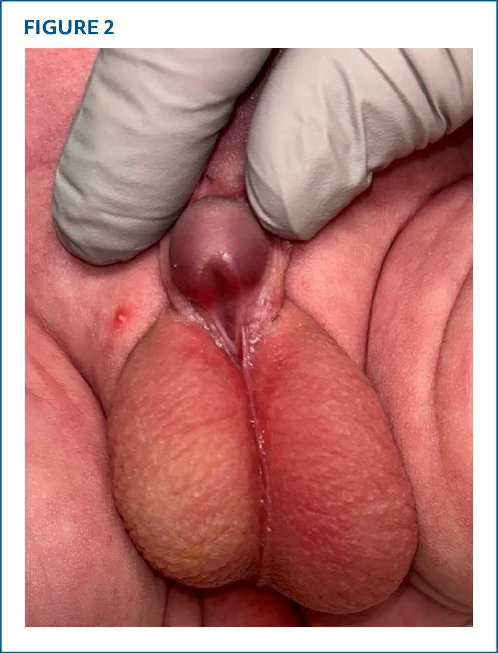
Physical examination revealed severe ventral chordee, dorsal hooded foreskin, adequate glans, and shortened penile length (Figures 1 and 2). The infant's testicles were descended bilaterally with a small hydrocele on the left side.
FIGURE 1
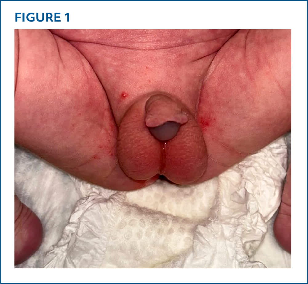
FIGURE 2
QUESTION
Based on the patient's history and physical examination, which one of the following is the most likely diagnosis?
- A. Differences of sexual development.
- B. Epispadias.
- C. Hypospadias.
- D. Megameatus with intact prepuce.
DISCUSSION
The answer is C: hypospadias. Hypospadias is one of the most common congenital anomalies in male infants, occurring in approximately 1 in 150 live male births.1 In this condition, the urethral meatus is abnormally located on the underside (ventral) aspect of the penis.2 These anomalies are commonly classified according to location: distal (subcoronal, glandular), midshaft (distal penile, proximal penile, midshaft), and proximal (scrotal, penoscrotal, perineal).3 The subcoronal location is the most common. Complications and repair complexity increase as the opening of the urethral meatus becomes more proximal.
This patient had proximal hypospadias in a penoscrotal location. Other features of hypospadias in this patient included hooded foreskin and ventral curvature of the penis (ventral chordee). Patients may also have cryptorchidism, inguinal hernias, underdeveloped fore-skin, and abnormal urine stream. Ultrasonography may reveal vesicoureteral reflux, ureteropelvic junction obstruction, pelvic or horseshoe kidney, crossed renal ectopia, or renal agenesis.4
Hypospadias should be identified on the newborn examination shortly after birth, and early detection is critical for management decisions. Circumcision should not be performed because the foreskin can be used for corrective surgery, which is usually performed at about 6 months of age.
Differences in sexual development can occur in conjunction with or have a similar presentation to hypospadias, including short penile shaft, hooded foreskin, chordee, and cryptorchidism. Other characteristics of differences in sexual development include ambiguous genitalia, absent or undescended testes, clitoromegaly micropenis, and labioscrotal folds. When unilateral or bilateral undescended testes are associated with hypospadias, pediatric endocrinology and pediatric urology should be consulted.5,6
Epispadias is the abnormal location of the urethral meatus on the dorsal aspect of the penis or on the abdomen. The embryologic origin is believed to be distinctly different from hypospadias, and epispadias is usually associated with other abnormalities.7
Megameatus with intact prepuce is a variant of hypospadias with a large, widened, patent urethral meatus located at or near the glans. In this patient, the observed central indentation could be confused for an elongated and widened meatus; however, no urine was observed coming from the central indentation, and it was not patent. Megameatus normally does not cause ventral chordee.8
SUMMARY TABLE
| Condition | Characteristics |
|---|---|
| Differences in sexual development | Short penile shaft, hooded foreskin, chordee, cryptorchidism, ambiguous genitalia, absent or undescended testes, clitoromegaly micropenis, labioscrotal folds |
| Epispadias | Urethral meatus located on the dorsal aspect of the penis or on the abdomen |
| Hypospadias | Urethral meatus located ventrally anywhere from glans to perineum |
| Megameatus with intact prepuce | Large, widened, patent meatus located at or near the glans |
Read the full article
Get immediate access, anytime, anywhere.
Choose a single article, issue, or full-access subscription.
Earn up to 14 CME credits per issue.