A 16-year-old male patient presented with a pruritic rash that had started on his face and spread rapidly to other areas of his body. He also had a fever and the sensation of something in his eye for the past week. The rash continued to worsen despite treatment with topical mupirocin and oral sulfamethoxazole/trimethoprim. The patient was a wrestler with a history of atopic dermatitis and acne.
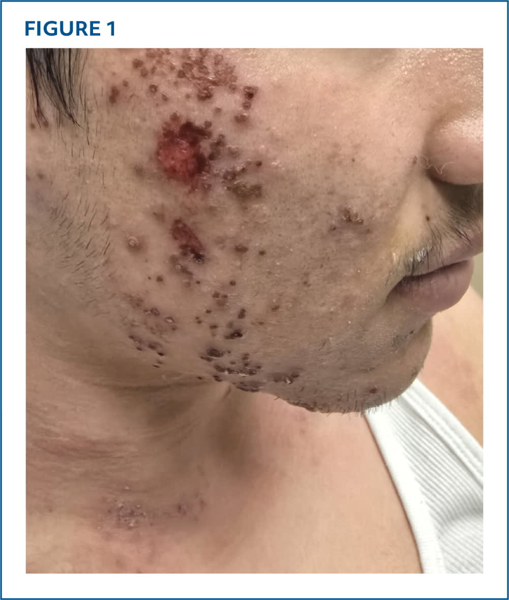
Physical examination revealed numerous coalescing punched-out erosions and papulovesicles on erythematous, xerotic bases. The lesions had hemorrhagic and honey-colored crusts with excoriations. The rash was extensive on the patient's forehead, temple, nose, chin, and right cheek (Figure 1). The surrounding skin showed perilesional erythema, mild edema, and satellite pustules. Cervical lymphadenopathy was present along with multiple erythematous lichenified plaques on his neck, shoulders, forearms, and anterior lower legs.
FIGURE 1
QUESTION
Based on the patient's history and physical examination, which one of the following is the most likely diagnosis?
- A. Allergic contact dermatitis.
- B. Bullous impetigo.
- C. Eczema herpeticum.
- D. Herpes gladiatorum.
- E. Primary varicella.
DISCUSSION
The answer is C: eczema herpeticum, a severe cutaneous infection caused by herpes simplex virus (HSV) type 1 or 2.1 The pathogenesis is complex and likely involves the interaction of impaired function of the epidermal barrier, dysregulated immune response, altered skin microbiota, and environmental factors.2 Eczema herpeticum typically occurs in individuals with skin conditions such as atopic, contact, or seborrheic dermatitis.1 It is often accompanied by fever, malaise, and lymphadenopathy. Clinically, it presents with an exacerbation of an associated dermatosis. Monomorphic, discrete, punched-out lesions may blister and rupture, resulting in erosions with overlying hemorrhagic crusts.3 The rash affects the face and trunk but can involve any area with underlying dermatosis. Eczema herpeticum is often misdiagnosed as eczematous dermatitis or impetigo. Diagnosis can be confirmed with polymerase chain reaction testing, Tzanck testing, direct fluorescent antibody testing, viral culture, or biopsy.1
Secondary infection with Staphylococcus aureus or Streptococcus pyogenes can lead to impetiginized lesions. Delay in treatment can result in HSV keratoconjunctivitis or keratitis (which can cause permanent vision loss), meningoencephalitis, gingivostomatitis, viremia, bacteremia, or death. With timely antiviral treatment, the mortality rate is 0.1%.2,3
Polymerase chain reaction analysis of this patient's ruptured papulovesicular lesions revealed HSV-1 infection. He was admitted for intravenous acyclovir therapy. Ophthalmologic assessment did not reveal corneal involvement. He was concurrently treated with cephalexin for bacterial superinfection and topical corticosteroids for atopic dermatitis. Within 2 weeks, his lesions were nearly resolved. Approximately 20% of patients experience recurrence and benefit from long-term viral suppression and optimized treatment of underlying skin conditions.1
Allergic contact dermatitis is a delayed (type IV) hypersensitivity reaction to haptens (small-molecule allergens that may evoke an immune response when they cross the skin barrier). On reexposure, a T-cell–mediated response is triggered. This condition typically presents with intense pruritus and an eczematous rash with demarcated borders.3
Bullous impetigo is caused by exfoliative toxins that occur with S aureus infection. It presents as small vesicles that enlarge into 1- to 2-cm superficial bullae that rupture to form a scaling rim with minimal surrounding erythema.3 Fever and lymphadenopathy are typically absent. Bullous impetigo can be self-limiting, but antibiotics may decrease duration and spread. Patients with immunodeficiency may develop staphylococcal scalded skin syndrome.4
Read the full article
Get immediate access, anytime, anywhere.
Choose a single article, issue, or full-access subscription.
Earn up to 14 CME credits per issue.