

Heel Pain: Diagnosis and Management

Nailah Adams Morancie, MD, MS
Landon Irvin, MD
Brian Z. Rayala, MD

American Family Physician. 2025;112(6):648-656.

Author disclosure: No relevant financial relationships.

This clinical content conforms to AAFP criteria for CME.

Heel pain accounts for 2 million office visits annually and is associated with higher body mass index, manual jobs, and a sedentary lifestyle. Retrocalcaneal bursitis is more common in women 40 to 60 years of age with Haglund deformity and a thickened Achilles tendon. Calcaneal apophysitis (Sever disease) is the most common cause of heel pain in active children and adolescents; in the outpatient setting, it accounts for up to 16% of musculoskeletal conditions in children. In os trigonum syndrome, athletes such as soccer players, gymnasts, and dancers who are engaged in repetitive plantar flexion commonly present with worsening posterior ankle pain and an antalgic gait. Risk factors for peroneal tendon injuries include corticosteroid injections into the peroneal sheath, use of fluoroquinolones, rheumatoid arthritis, hyperparathyroidism, and diabetes. Magnetic resonance imaging is the most accurate test for the diagnosis of most causes of heel pain, but ultrasonography is being used more often as it becomes more readily available at the point of care. Most patients with heel pain improve with conservative treatment, and surgical management should be reserved for recalcitrant cases. Pain catastrophizing and kinesiophobia are associated with diminished foot function and poorer prognosis.

Heel pain accounts for 2 million office visits annually and is associated with higher body mass index, manual jobs, and a sedentary lifestyle.1 Evaluation and management of heel pain are based on the location of pain (Figure 12). Mechanical etiologies, such as Achilles tendinopathy, are common causes of heel pain (Table 12). However, neurologic, arthritic, oncologic, infectious, and traumatic etiologies should be considered. Figure 2 provides a practical approach for the diagnosis of heel pain.2

SORT: KEY RECOMMENDATIONS FOR PRACTICE
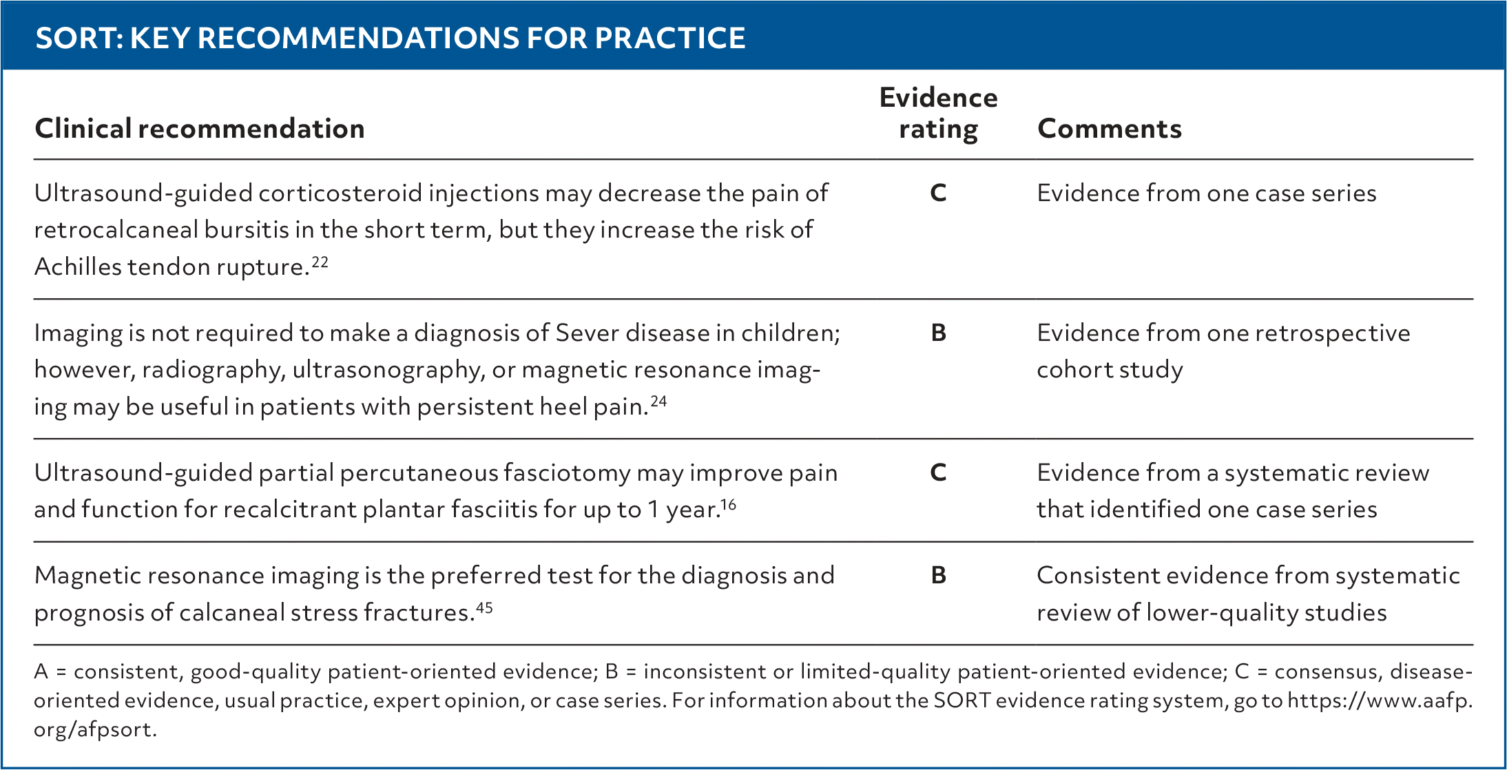
Clinical recommendationEvidence ratingComments
Ultrasound-guided corticosteroid injections may decrease the pain of retrocalcaneal bursitis in the short term, but they increase the risk of Achilles tendon rupture.22 CEvidence from one case series
Imaging is not required to make a diagnosis of Sever disease in children; however, radiography, ultrasonography, or magnetic resonance imaging may be useful in patients with persistent heel pain.24 BEvidence from one retrospective cohort study
Ultrasound-guided partial percutaneous fasciotomy may improve pain and function for recalcitrant plantar fasciitis for up to 1 year.16 CEvidence from a systematic review that identified one case series
Magnetic resonance imaging is the preferred test for the diagnosis and prognosis of calcaneal stress fractures.45 BConsistent evidence from systematic review of lower-quality studies

A = consistent, good-quality patient-oriented evidence; B = inconsistent or limited-quality patient-oriented evidence; C = consensus, disease-oriented evidence, usual practice, expert opinion, or case series. For information about the SORT evidence rating system, go to https://www.aafp.org/afpsort.

FIGURE 1.
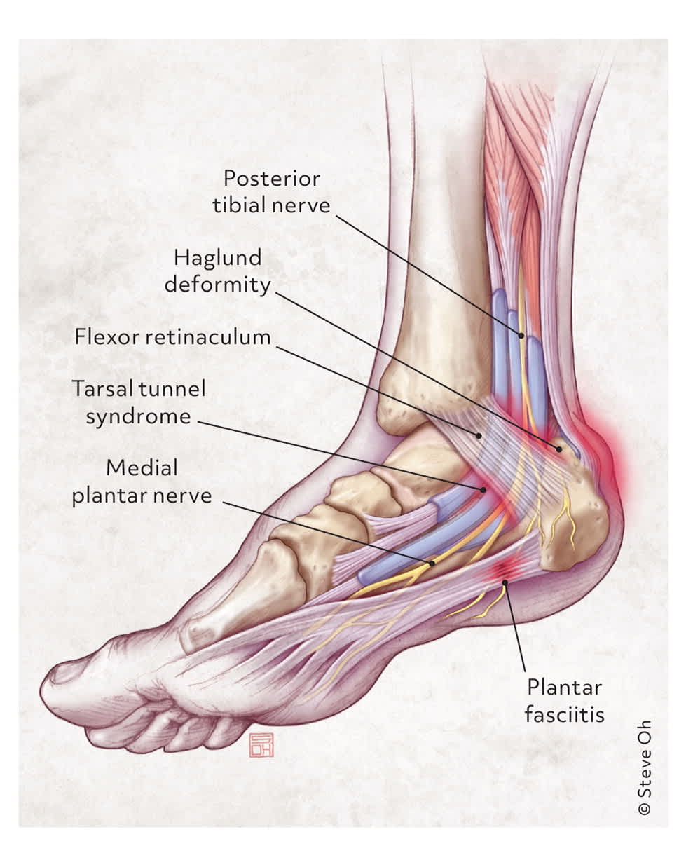
Heel pain and corresponding anatomy.

Illustration © Steve Oh

Adapted with permission from Tu P, Bytomski JR. Diagnosis of heel pain. Am Fam Physician. 2011;84(8):912.

TABLE 1. Mechanical Causes of Heel Pain by Location
EtiologyClinical featuresInitial treatment
Posterior
Achilles tendinopathyAchy, sharp pain with tenderness along the tendon
Worsens with increased activity
Occasional palpable prominence from tendon thickening
Worsens with passive dorsiflexion
Activity modification, rest and weight-bearing as tolerated, decrease pressure to affected area, heel lifts or orthotics, NSAIDs or analgesics, deep friction massage, ankle bracing, and eccentric exercises

Haglund deformityPain at the superior aspect of the posterior calcaneus
Radiography may show early deformities
Decrease pressure to affected area and NSAIDs or analgesics

Retrocalcaneal bursitisPain, erythema, and swelling around the Achilles tendon
Tenderness on direct palpation
Decrease pressure to affected area, NSAIDs or analgesics, and corticosteroid injections (preferably ultrasound guided)

Calcaneal apophysitis (Sever disease)Pain in adolescents that worsens with increased activity or during growth spurt
Tenderness at Achilles insertion
Pain with passive dorsiflexion and mediolateral calcaneal compression
Activity modification, NSAIDs or analgesics, ice, stretching and strengthening exercises, and orthotics or shoe modifications

Inferior
Calcaneal stress fractureCommonly occurs following an increase in activity level or a change to harder walking surfaces
Pain with activity that progressively worsens to pain at rest
Diagnosed with imaging
Activity modification with occasional non–weight-bearing activity, heel pads, walking boots, crutches, or a controlled ankle motion walker

Heel pad syndromeDeep, bruise-like pain and tenderness at middle of heelDecrease pressure to affected area, NSAIDs or analgesics, heel cups, taping, and proper footwear

Heel spursRadiographic findings at site of painDecrease pressure to affected area

Nerve entrapment (medial or lateral plantar nerve, nerve to abductor digiti minimi)Sensations of burning, tingling, or numbness
Occasionally preceded by increased activity or trauma
Decrease pressure to affected area, NSAIDs or analgesics, ice, and stretching exercises

NeuromaSensations of burning or tingling
Painful lump with palpation
Decrease pressure to affected area

Plantar fasciitisPain with first steps after prolonged rest
Tenderness on medial calcaneal tuberosity and plantar fascia
Rest and activity modification, stretching and strengthening exercises, NSAIDs or analgesics, ice massage, and arch support

Midfoot (medial)
Flexor digitorum longus tendinopathyTenderness posterior to medial malleolus and obliquely across sole of foot to base of distal phalanges of lateral toesActivity modification, decrease pressure to affected area, NSAIDs or analgesics, and eccentric exercises

Flexor hallucis longus tendinopathyTenderness posterior to medial malleolus and on plantar surface of great toeActivity modification, decrease pressure to affected area, NSAIDs or analgesics, and eccentric exercises

Posterior tibialis tendinopathyTenderness at navicular and medial cuneiformActivity modification, decrease pressure to affected area, NSAIDs or analgesics, and eccentric exercises

Tarsal tunnel syndromeBurning, tingling, or shooting pain and numbness in posteromedial ankle and heel (may extend to toes) that worsens with standing and activity
Positive Tinel sign
Muscle atrophy in severe cases
Activity modification, orthotics, NSAIDs, neuromodulator therapy, and corticosteroid injections

Midfoot (lateral)
Peroneal tendinopathyTenderness in lateral calcaneus along path to base of fifth metatarsalActivity modification, decrease pressure to affected area, NSAIDs or analgesics, and eccentric exercises

Sinus tarsi syndromePain in lateral calcaneus and ankle
Feeling of foot or ankle instability
Worse after exercise or on uneven surfaces
May have history of repeated ankle sprains
Orthotics, ice massage, NSAIDs or analgesics, physical therapy (eg, balance/proprioception, strengthening exercises), and corticosteroid injections

NSAID = nonsteroidal anti-inflammatory drug.

Adapted with permission from Tu P, Bytomski JR. Diagnosis of heel pain. Am Fam Physician. 2011;84(8):910–911.

NAILAH ADAMS MORANCIE, MD, MS, CAQSM, FAAFP, is an assistant professor in the Department of Family Medicine at the University of North Carolina at Chapel Hill.

LANDON IRVIN, MD, CAQSM, is an assistant professor in the Department of Family Medicine at the University of North Carolina at Chapel Hill.

BRIAN Z. RAYALA, MD, FAAFP, is a professor in the Department of Family Medicine at the University of North Carolina at Chapel Hill.

Address correspondence to Nailah Adams Morancie, MD, MS, CAQSM, FAAFP, at nailah_adams@med.unc.edu.

Author disclosure: No relevant financial relationships.

  1. 1.Chatterton BD, Muller S, Roddy E. Epidemiology of posterior heel pain in the general population: cross-sectional findings from the clinical assessment study of the foot. Arthritis Care Res (Hoboken). 2015;67(7):996-1003.
  2. 2.Tu P, Bytomski JR. Diagnosis of heel pain. Am Fam Physician. 2011;84(8):909-916.
  3. 3.Cotchett M, Frescos N, Whittaker GA, et al. Psychological factors associated with foot and ankle pain: a mixed methods systematic review. J Foot Ankle Res. 2022;15(1):10.
  4. 4.Schwieterman B, Haas D, Columber K, et al. Diagnostic accuracy of physical examination tests of the ankle/foot complex: a systematic review. Int J Sports Phys Ther. 2013;8(4):416-426.
  5. 5.Perhamre S, Lazowska D, Papageorgiou S, et al. Sever’s injury: a clinical diagnosis. J Am Podiatr Med Assoc. 2013;103(5):361-368.
  6. 6.De Garceau D, Dean D, Requejo SM, et al. The association between diagnosis of plantar fasciitis and Windlass test results. Foot Ankle Int. 2003;24(3):251-255.
  7. 7.Bubra PS, Keighley G, Rateesh S, et al. Posterior tibial tendon dysfunction: an overlooked cause of foot deformity. J Family Med Prim Care. 2015;4(1):26-29.
  8. 8.Yasui Y, Tonogai I, Rosenbaum AJ, et al. The risk of Achilles tendon rupture in the patients with Achilles tendinopathy: healthcare database analysis in the United States. BioMed Res Int. ;2017:7021862.
  9. 9.Patch DA, Andrews NA, Scheinberg M, et al. Achilles tendon disorders: an overview of diagnosis and conservative treatment. JAAPA. 2023;36(10):1-8.
  10. 10.Tonarelli JM, Mabry LM, Ross MD. Diagnostic imaging of an Achilles tendon rupture. J Orthop Sports Phys Ther. 2011;41(11):904.
  11. 11.Fenech M, Ajjikuttira A, Edwards H. Ultrasound assessment of acute Achilles tendon rupture and measurement of the tendon gap. Australas J Ultrasound Med. 2024;27(2):106-119.
  12. 12.Chiodo CP, Glazebrook M, Bluman EM, et al. American Academy of Orthopaedic Surgeons clinical practice guideline on treatment of Achilles tendon rupture. J Bone Joint Surg Am. 2010;92(14):2466-2468.
  13. 13.Myhrvold SB, Brouwer EF, Andresen TKM, et al. Nonoperative or surgical treatment of acute Achilles’ tendon rupture. N Engl J Med. 2022;386(15):1409-1420.
  14. 14.Moraes VY, Lenza M, Tamaoki MJ, et al. Platelet-rich therapies for musculoskeletal soft tissue injuries. Cochrane Database Syst Rev. 2014(4):CD010071.
  15. 15.Kearney RS, Parsons N, Metcalfe D, et al. Injection therapies for Achilles tendinopathy. Cochrane Database Syst Rev. 2015(5):CD010960.
  16. 16.Vajapey S, Ghenbot S, Baria MR, et al. Utility of percutaneous ultrasonic tenotomy for tendinopathies: a systematic review. Sports Health. 2021;13(3):258-264.
  17. 17.Laboute E, Thoreux P, Beldame J, et al. Re-ruptures and early outcomes after surgical repair of acute Achilles tendon ruptures: prospective, comparative multicenter study. Int Orthop. 2023;47(7):1855-1861.
  18. 18.Wnuk-Scardaccione A, Mizia E, Zawojska K, et al. Surface shape of the calcaneal tuberosity and the occurrence of retrocalcaneal bursitis among runners. Int J Environ Res Public Health. 2021;18(6):2860.
  19. 19.Uquillas CA, Guss MS, Ryan DJ, et al. Everything Achilles: knowledge update and current concepts in management: AAOS exhibit selection. J Bone Joint Surg Am. 2015;97(14):1187-1195.
  20. 20.Olivieri I, Barozzi L, Padula A, et al. Retrocalcaneal bursitis in spondyloarthropathy: assessment by ultrasonography and magnetic resonance imaging. J Rheumatol. 1998;25(7):1352-1357.
  21. 21.Maffulli N, Saxena A, Wagner E, et al. Achilles insertional tendinopathy: state of the art. J ISAKOS. 2019;4(1):48-57.
  22. 22.Boone SL, Uzor R, Walter E, et al. Safety and efficacy of image-guided retrocalcaneal bursa corticosteroid injection for the treatment of retrocalcaneal bursitis. Skeletal Radiol. 2021;50(12):2471-2482.
  23. 23.Wiegerinck JI, Vroemen JC, van Dongen TH, et al. The posterior impingement view: an alternative conventional projection to detect bony posterior ankle impingement. Arthroscopy. 2014;30(10):1311-1316.
  24. 24.Belikan P, Färber LC, Abel F, et al. Incidence of calcaneal apophysitis (Sever’s disease) and return-to-play in adolescent athletes of a German youth soccer academy: a retrospective study of 10 years. J Orthop Surg Res. 2022;17(1):83.
  25. 25.Duong MM, Nicholson AD, Li SQ, et al. Relationship between Sever disease and skeletal maturity. J Pediatr Orthop. 2020;40(2):93-96.
  26. 26.Sweeney EA, Little CC, Wilson JC, et al. Comparison of braces for treatment of Sever’s disease (calcaneal apophysitis) in barefoot athletes: a randomized clinical trial. J Athl Train. 2023;58(5):437-444.
  27. 27.Hernandez-Lucas P, Leirós-Rodríguez R, García-Liñeira J, et al. Conservative treatment of Sever’s disease: a systematic review. J Clin Med. 2024;13(5):1391.
  28. 28.Knapik DM, Guraya SS, Jones JA, et al. Incidence and fusion of os trigonum in a health pediatric population. J Pediatr Orthop. 2019;39(9):e718-e721.
  29. 29.McAlister JE, Urooj U. Os trigonum syndrome. Clin Podiatr Med Surg. 2021;38(2):279-290.
  30. 30.Ishibashi MA, Doyle MD, Krcal CE. Posterior ankle impingement syndrome. Clin Podiatr Med Surg. 2023;40(1):209-222.
  31. 31.Robinson P, Bollen SR. Posterior ankle impingement in professional soccer players: effectiveness of sonographically guided therapy. AJR Am J Roentgenol. 2006;187(1):W53-W58.
  32. 32.Kakouris N, Yener N, Fong DTP. A systematic review of running-related musculoskeletal injuries in runners. J Sport Health Sci. 2021;10(5):513-522.
  33. 33.Khammas ASA, Mahmud R, Hassan HA, et al. An assessment of plantar fascia with ultrasound findings in patients with plantar fasciitis: a systematic review. J Ultrasound. 2023;26(1):13-38.
  34. 34.Albano D, Messina C, Gitto S, et al. Shear-wave elastography of the plantar fascia: a systematic review and meta-analysis. J Ultrasound. 2023;26(1):59-64.
  35. 35.Chundru U, Liebeskind A, Seidelmann F, et al. Plantar fasciitis and calcaneal spur formation are associated with abductor digiti minimi atrophy on MRI of the foot. Skeletal Radiol. 2008;37(6):505-510.
  36. 36.Seligman DAR, Dawson D, Streiner DL, et al. Treating heel pain in adults: a randomized controlled trial of hard vs modified soft custom orthotics and heel pads. Arch Phys Med Rehabil. 2021;102(3):363-370.
  37. 37.Reinstein M, Weisman A, Masharawi Y. Barefoot walking is beneficial for individuals with persistent plantar heel pain: a single-blind randomized controlled trial. Ann Phys Rehabil Med. 2024;67(2):101786.
  38. 38.Elizondo-Rodríguez J, Simental-Mendía M, Peña-Martínez V, et al. Comparison of botulinum toxin A, corticosteroid, and anesthetic injection for plantar fasciitis. Foot Ankle Int. 2021;42(3):305-313.
  39. 39.David JA, Sankarapandian V, Christopher PR, et al. Injected corticosteroids for treating plantar heel pain in adults. Cochrane Database Syst Rev. 2017(6):CD009348.
  40. 40.Naruseviciute D, Kubilius R. The effect of high-intensity versus low-level laser therapy in the management of plantar fasciitis: randomized participant blind controlled trial. Clin Rehabil. 2020;34(8):1072-1082.
  41. 41.Italiano J, Bitterman AD. Diagnosis and management of calcaneal stress fractures. Radiol Technol. 2021;93(2):177-194.
  42. 42.Walsh TP, Vasudeva V, Sampang K, et al. Psychological dysfunction associated with calcaneal fractures. Injury. 2021;52(8):2475-2478.
  43. 43.Vosoughi AR, Borazjani R, Ghasemi N, et al. Different types and epidemiological patterns of calcaneal fractures based on reviewing CT images of 957 fractures. Foot Ankle Surg. 2022;28(1):88-92.
  44. 44.Mountjoy M, Ackerman KE, Bailey DM, et al. 2023 International Olympic Committee’s (IOC) consensus statement on relative energy deficiency in sport (REDs). Br J Sports Med. 2023;57(17):1073-1097.
  45. 45.Hoenig T, Tenforde AS, Strahl A, et al. Does magnetic resonance imaging grading correlate with return to sports after bone stress injuries? A systematic review and meta-analysis. Am J Sports Med. 2022;50(3):834-844.
  46. 46.Ferkel E, Davis WH, Ellington JK. Entrapment neuropathies of the foot and ankle. Clin Sports Med. 2015;34(4):791-801.
  47. 47.Kiel J, Kaiser K. Tarsal tunnel syndrome. StatPearls. Accessed June 5, 2024. https://www-ncbi-nlm-nih-gov.libproxy.lib.unc.edu/books/NBK513273/
  48. 48.Urits I, Smoots D, Franscioni H, et al. Injection techniques for common chronic pain conditions of the foot: a comprehensive review. Pain Ther. 2020;9(1):145-160.
  49. 49.Rehman M, Duarte Silva F, Chhabra A. Diagnostic efficacy of posterior tibialis tendon dysfunction: a systematic review of literature. Eur Radiol. 2024;34(5):3513-3523.
  50. 50.Molloy R, Tisdel C. Failed treatment of peroneal tendon injuries. Foot Ankle Clin. 2003;8(1):115-129.
  51. 51.Willegger M, Hirtler L, Schwarz GM, et al. Peroneal tendon pathologies: from the diagnosis to treatment. Orthopade. 2021;50(7):589-604.
  52. 52.Pesquer L, Guillo S, Poussange N, et al. Dynamic ultrasound of peroneal tendon instability. Br J Radiol. 2016;89(1063):20150958.
  53. 53.Lamm BM, Myers DT, Dombek M, et al. Magnetic resonance imaging and surgical correlation of peroneus brevis tears. J Foot Ankle Surg. 2004;43(1):30-36.
  54. 54.Tu P. Heel pain: diagnosis and treatment. Am Fam Physician. 2018;97(2):86-93.
  55. 55.Aldridge T. Diagnosing heel pain in adults. Am Fam Physician. 2004;70(2):332-338.

Copyright © 2026 by the American Academy of Family Physicians.

This content is owned by the AAFP. A person viewing it online may make one printout of the material and may use that printout only for his or her personal, non-commercial reference. This material may not otherwise be downloaded, copied, printed, stored, transmitted or reproduced in any medium, whether now known or later invented, except as authorized in writing by the AAFP. See permissions for copyright questions and/or permission requests.