An active 81-year-old woman was evaluated 3 days after injuring her right hand while playing pickleball. After hitting the ball with extra force, she had immediately felt pain over the dorsum of her right hand. She then noticed progressive swelling of the affected area over the next 2 days.
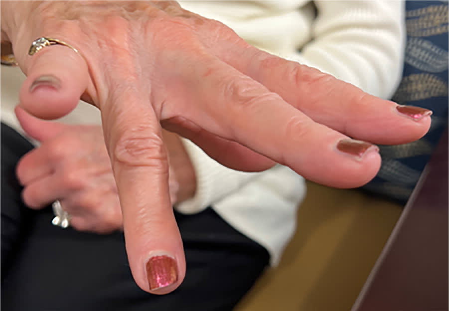
Examination revealed swelling over the metacarpal of her right fourth finger, and she had difficulty extending that finger (Figure 1).
FIGURE 1
QUESTION
Based on the patient's history and physical examination, which one of the following is the most likely diagnosis?
- A. Central extensor tendon injury with boutonnière deformity.
- B. Distal extensor tendon rupture with dorsal avulsion fracture (mallet finger).
- C. Dupuytren contracture.
- D. Extensor digitorum tendon rupture.
- E. Flexor digitorum profundus avulsion fracture.
DISCUSSION
The correct answer is D: extensor digitorum tendon rupture, which is characterized by a reduced ability to extend the affected finger at the metacarpophalangeal joint. Extensor tendon injuries of the hand are common, with an incidence of 14 per 100,000 person-years. They occur twice as often as flexor injuries of the fingers. Extensor tendon injuries are categorized into eight zones, with odd-numbered zones over joints and even-numbered zones between them.1 This patient's injury occurred in zone six, over the metacarpal bone of the fourth finger. Extensor tendon injuries most commonly involve the index finger or thumb; injuries in zone six are less common, accounting for only 5% of hand tendon injuries.2
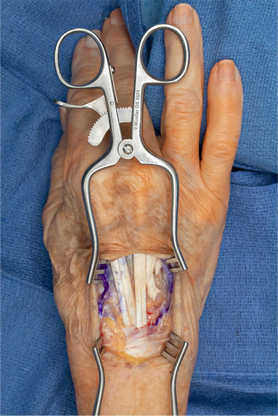
This patient was fitted with a wrist splint and referred to a hand surgeon. Radiograph findings were negative, but magnetic resonance imaging showed extensor digitorum tendon injury. During surgery, the patient was found to have significant synovitis and rupture of the extensor digitorum tendons of the fourth and fifth right fingers (Figure 2). The extensor digiti minimi tendon was intact. The ruptured tendons were sewn together using a running, locking suture and horizontal mattress sutures. This technique results in less tendon loss and improved strength compared with alternative techniques.3 Postoperatively, the patient completed a course of dynamic splinting and range-of-motion therapy over 12 weeks. Early active-motion protocols with dynamic extension orthoses provide better total active motion and final grip strength than those with static orthoses.4
FIGURE 2
Boutonnière deformity is characterized by a finger flexed at the proximal interphalangeal (PIP) joint and hyperextended at the distal interphalangeal (DIP) joint. This condition results from a central slip rupture of the extensor tendon at the PIP joint. Common causes of boutonnière deformity are trauma, rheumatoid arthritis, and Dupuytren contracture. The Elson test can be used to evaluate for possible tearing of the central slip before the deformity becomes evident. In this test, the patient bends the PIP joint to a 90-degree angle over the edge of a table and then attempts to extend the middle phalanx of the finger against the examiner's resistance. When the central slip is intact (a negative test result), the examiner feels extension at the PIP joint as the finger extends.5,6
Mallet finger is a distal extensor tendon rupture accompanied by a dorsal avulsion fracture, usually caused by forced flexion at the DIP joint. This results in the inability to actively extend the DIP joint, with the injured finger remaining flexed.7,8
Read the full article
Get immediate access, anytime, anywhere.
Choose a single article, issue, or full-access subscription.
Earn up to 14 CME credits per issue.