To the Editor: Soft tissue masses commonly present in family medicine and range from benign lipomas to high-grade soft tissue sarcomas. A subset of soft tissue sarcomas includes a spindle cell sarcoma, which is a rare connective tissue tumor that originates in the layers of tissue found below the skin.1 Spindle cell sarcomas can be found on any part of the body, but most occur on the trunk and extremities.2 Surgical resection is the mainstay of treatment for spindle cell sarcomas, but treatment can also include a combination of surgery, chemotherapy, and radiation.1
A previously healthy 45-year-old woman had a mechanical fall resulting in ecchymosis of the left eye and left thigh pain. Two weeks after the fall, she noticed a small bump on her left thigh. The patient made an appointment with her family physician. The initial evaluation included ultrasonography, but ultimately magnetic resonance imaging of the left leg revealed a large collection in the lateral thigh consistent with a hematoma. A neoplastic process could not be ruled out. Over the next month, the mass on the patient's thigh continued to grow. A general surgeon ordered a computed tomography angiogram of the pelvis and bilateral lower extremities that showed a moderate-sized fluid collection within the soft tissues anterior to the left hip, suggestive of a chronic hematoma. The patient was referred to surgical oncology and had a core needle biopsy of the mass done. Biopsy results showed completely necrotic cells with no visible tumor cells present.
Five days after the biopsy, the patient presented to the hospital with worsening pain and erythema over the anterior left thigh. Magnetic resonance imaging of her left hip revealed an interval increase in the size of the complex mass in the left thigh (Figure 1). The patient was taken to the operating room by general surgery for excision of the thigh mass and drain placement. During the surgery, a large, encapsulated mass measuring 17 cm (6.7 in) by 17 cm, with serous mucinous fluid was removed with the capsule intact. The pathology of the encapsulated mass revealed an unclassified high-grade spindle cell sarcoma which had been mimicking an intramuscular hematoma. The patient's postoperative course was unremarkable. The diagnosis of sarcoma is often delayed because of its rarity and nonspecific presenting symptoms.3 This case illustrates the importance of including sarcoma in the differential diagnosis in patients presenting with a soft tissue mass and hematoma.
FIGURE 1.
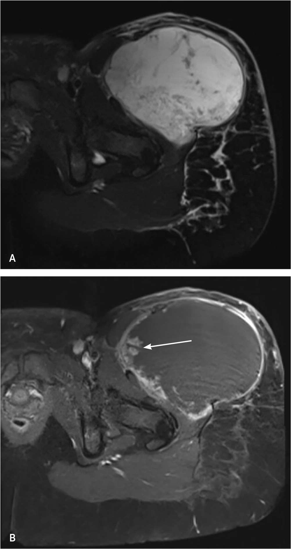
(A) Axial T2-weighted fat-suppressed magnetic resonance imaging (MRI) scan, and (B) axial T1-weighted fat-saturation MRI scan post-contrast showing a large complex cystic mass in the left thigh with areas of nodular enhancement along its margins (arrow).
