To the Editor: A healthy 13-month-old was brought to the clinic for his one-year well-child examination. After an uneventful prenatal course, he was born at term to a 33-year-old patient via normal spontaneous vaginal delivery. The Apgar scores were 8 and 9 at one and five minutes, respectively. The infant has attended all recommended well-child examinations, and his vaccinations are up-to-date. His developmental milestones have been age-appropriate. During this visit, his weight was in the 98th percentile, height in the 90th percentile, and head circumference greater than the 99th percentile. On physical examination, his head appeared disproportionately large for his body. The anterior fontanelle was soft and flat, and his gait was normal.
A head ultrasound showed dilation of the right lateral ventricle with a right-sided deviation of brain matter. Magnetic resonance imaging showed a 2.5-cm lobulated intraventricular mass within the right foramen of Monro extending into the third lateral ventricle and the frontal horn of the right lateral ventricle with a right to left shift of approximately 13.5 mm (Figures 1A and 1B). Surgical excision of the tumor was performed the next day. Pathology confirmed a choroid plexus papilloma. Repeat magnetic resonance imaging showed a successful choroid mass resection with interval improvement in the right lateral ventricle hydrocephalus (Figures 1C and 1D).
FIGURE 1.
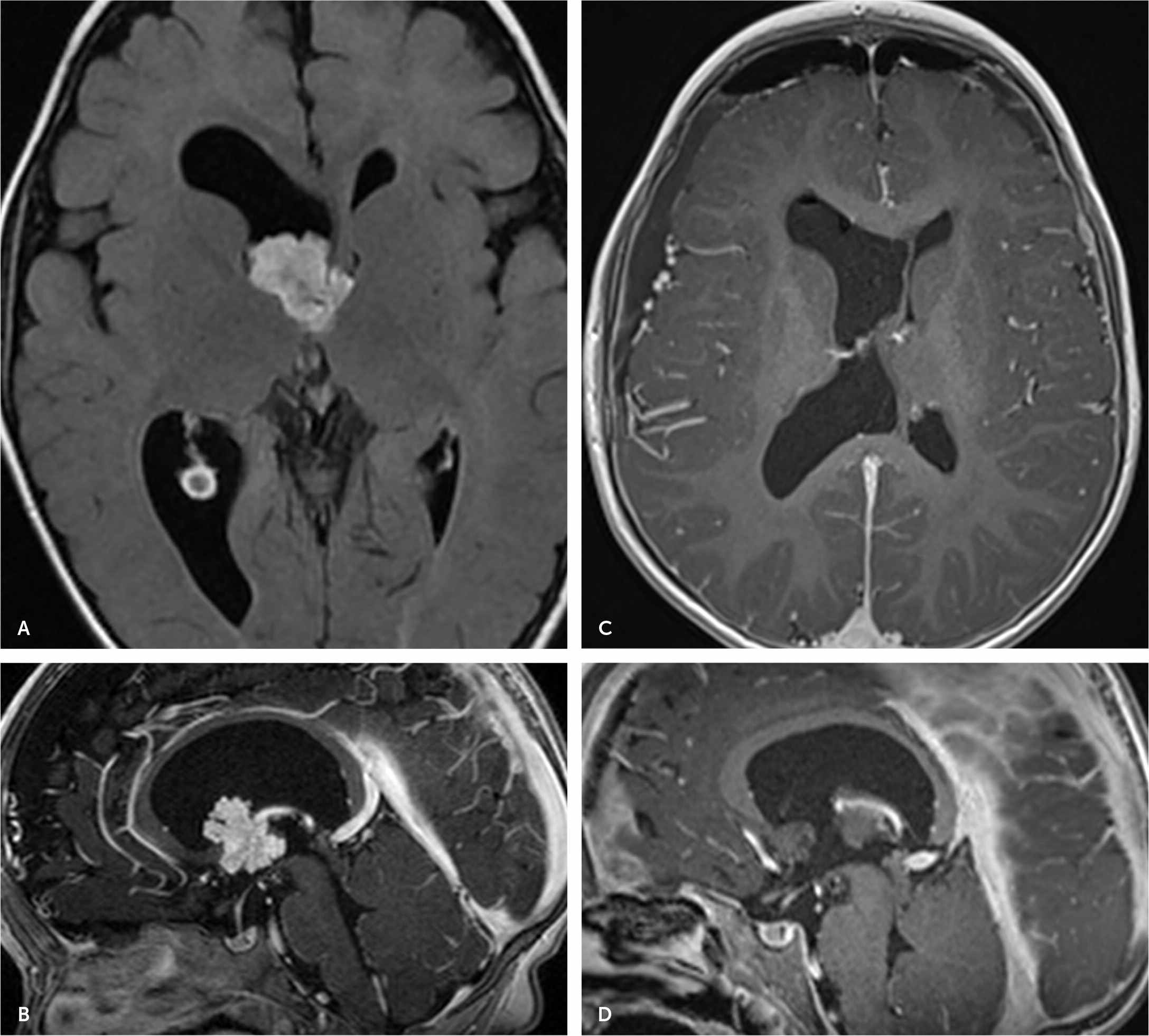
(A, B) Magnetic resonance imaging (MRI) conducted before surgery showing minimally lobulated intraventricular mass centered at the right foramen of Monro, extending into the third lateral ventricle and the frontal horn of the right lateral ventricle, resulting in dilation of the right lateral ventricle. (C, D) Repeat MRI after surgery.
Choroid plexus papillomas are rare central nervous system tumors that are diagnosed at a median age of four years.1 They are generally benign with an overall survival rate of 90% to 100% but may present with symptoms of hydrocephalus, seizures, or focal neurologic deficits.1 This case report emphasizes the importance of clinical observation and a thorough physical examination in making a timely diagnosis, and collaboration between family physicians and pediatric neurosurgeons to provide the best care for our patients.
