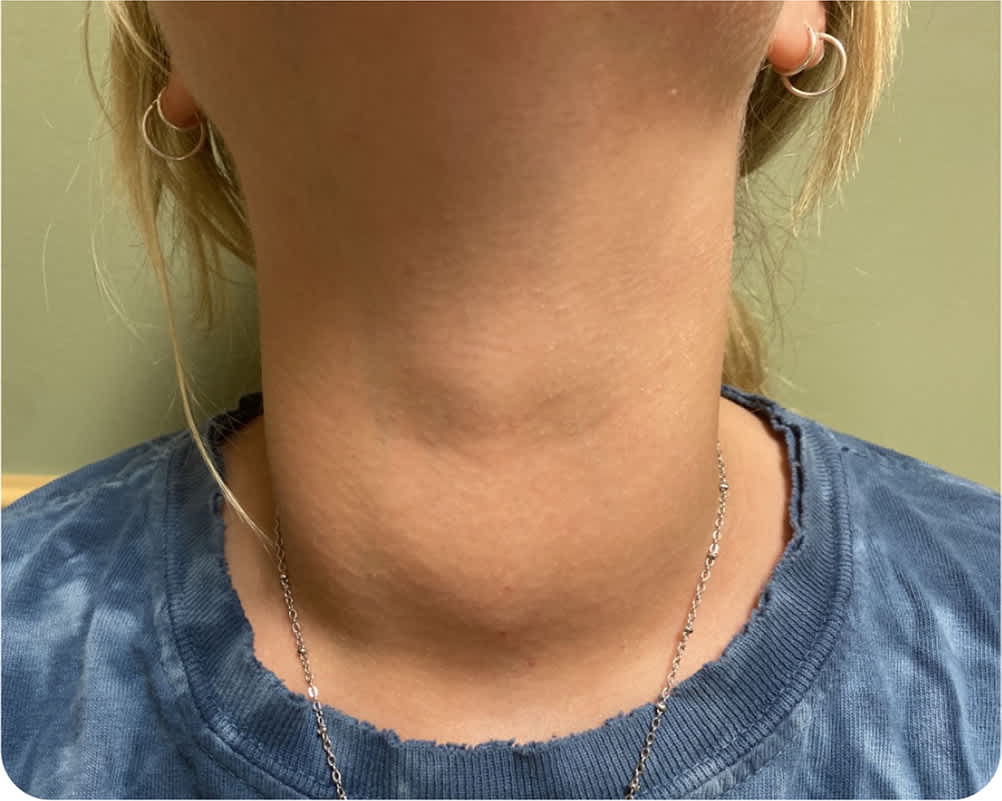
A 27-year-old woman presented with a mass on the front of her neck that had enlarged over the past few months. The mass was not tender or painful, but the patient had occasional dysphagia. She reported new symptoms, including irregular menses, heat intolerance, and increased anxiety and appetite. She was not taking any medications.
Physical examination revealed tachycardia with a heart rate of 120 beats per minute and a goiter that was diffusely enlarged and nontender (Figure 1). Her thyroid-stimulating hormone (TSH) level was less than 0.007 mIU per L, and her free thyroxine (T4) level was more than 8 ng per dL (102.97 pmol per L).
FIGURE 1
Question
Based on the patient's history and physical examination, which one of the following is the most likely diagnosis?
- A. de Quervain thyroiditis.
- B. Graves disease.
- C. Hashimoto thyroiditis.
- D. Toxic thyroid adenoma.
Discussion
The answer is B: Graves disease. This autoimmune disorder is the most common cause of hyperthyroidism in the United States, accounting for 60% to 80% of cases. It occurs in 20 to 50 out of 100,000 adults and is more common in women than men.1 Risk factors include a family history of the condition, other autoimmune diseases, stress, smoking, infection, and iodine exposure.2 In Graves disease, thyroid-stimulating antibodies agonize the TSH receptor. This leads to overproduction of the T4 hormone, causing hyperplasia of the thyroid gland with a resultant goiter that is diffuse and nontender.2
The goiter can also have bruits overlying the thyroid gland. Graves disease is typically diagnosed in younger patients but can also present at an older age. Younger patients tend to present with the classic symptoms of hyperthyroidism (i.e., heat intolerance, sweating, fatigue, palpitations, tremors, and anxiety).1 Older patients may have nonspecific symptoms of fatigue, weight loss, and atrial fibrillation. Typical physical examination findings include tachycardia, hypertension, muscle weakness, and hair loss.1 Laboratory findings are characterized by a low TSH level and an elevated total triiodothyronine (T3)/free T4 ratio. A radioactive iodine uptake scan of the thyroid shows diffusely high uptake.3
If untreated, T4 overproduction can have two major physical manifestations, ocular and musculoskeletal. Ocular manifestations can include exophthalmos, which presents with proptosis, dry eye, and ocular discomfort.1 Exophthalmos is caused by trapped water due to overproduction and deposition of glycosaminoglycans by TSH receptor–laden fibroblasts. This can lead to periorbital edema and irreversible fibrosis.2 Musculoskeletal complications include pretibial myxedema and thyroid acropachy. The musculoskeletal manifestations typically occur in patients with exophthalmos. Acropachy only occurs after development of dermopathy.1
De Quervain thyroiditis may present clinically as hyperthyroidism, hypothyroidism, or euthyroidism. Patients may recall a preliminary insult, such as a viral illness. Physical examination shows a tender goiter, which is often associated with painful neck movements.4
Hashimoto thyroiditis is the leading cause of hypothyroidism in populations with sufficient iodine intake. In this condition, TSH level is elevated, and T4 level is decreased. The destructive phase typically occurs over years, with periods of euthyroid readings.5
A toxic thyroid adenoma typically presents with hyperthyroid symptoms and a decreased TSH level and an elevated T4 level. Physical examination of the gland may reveal a palpable solitary thyroid nodule or area of enlargement.3
SUMMARY TABLE
| Condition | Characteristics |
|---|---|
| de Quervain thyroiditis | Laboratory values can vary; associated with prelimnary insult, such as a viral illness; tender goiter |
| Graves disease | Most common cause of hyperthyroidism in the United States; low TSH level, elevated total T3/free T4 ratio; associated with a nontender goiter |
| Hashimoto thyroiditis | Leading cause of hypothyroidism in iodine-sufficient areas; elevated TSH level and decreased T4 level; temporary hyperthyroid state and periods of euthyroid readings |
| Toxic thyroid adenoma | Hyperthyroid symptoms; low TSH level, elevated free T4 level; solitary thyroid nodule or enlarged area |
TSH = thyroid-stimulating hormone; T3 = triiodothyronine; T4 = thyroxine.
