

A 30-year-old man presented to the emergency department after the sole of his foot impacted a rock in a shallow pool of water. He reported experiencing immediate pain in the mid- and forefoot. He was able to walk, but weight-bearing caused pain. The patient had no previous injuries or surgeries to the foot.
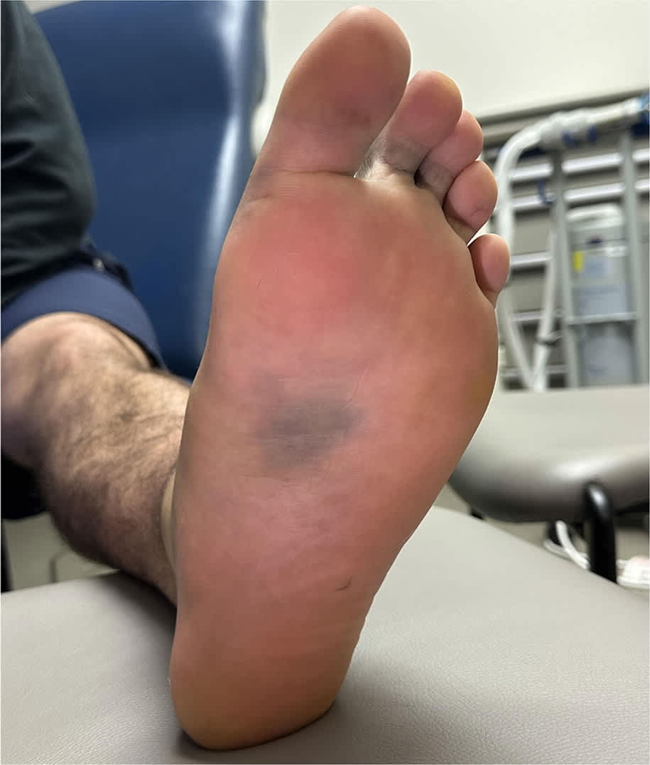
Evaluation revealed swelling and erythema on the plantar midfoot. A faint ecchymosis was developing on the surrounding plantar surface. The dorsal and plantar midfoot regions were tender, but no point tenderness was elicited over the lateral or medial malleoli, base of the fifth metatarsal bone, cuboid bone, navicular bone, calcaneus, or first metatarsophalangeal joint. Non–weight-bearing radiography showed no fractures of the foot or ankle. The patient was discharged with a controlled ankle motion boot and a follow-up appointment with his primary care physician.
After 3 days, he reported that weight-bearing caused severe pain in the midfoot. The midfoot was tender and had extensive ecchymosis on the plantar surface (Figure 1). Weight-bearing radiography showed widening between the intermediate and lateral cuneiform bones, the second and third metatarsal heads, and the first and second metatarsal bases. No fractures or other abnormalities were identified.
FIGURE 1
QUESTION
Based on the patient’s history and physical examination, which one of the following is the most likely diagnosis?
- A. Cuboid subluxation.
- B. Fifth metatarsal fracture.
- C. Navicular fracture.
- D. Plantar fascia rupture.
- E. Tarsometatarsal joint complex injury.
DISCUSSION
The answer is E: tarsometatarsal joint complex injury. The tarsometatarsal joint, also known as the Lisfranc joint, plays a key role in maintaining the foot arch. The joint complex is composed of the tarsometatarsal, intertarsal, and proximal intermetatarsal joints and supported by multiple ligaments. Acute traumatic injuries are usually high-energy mechanisms involving direct blunt trauma to the midfoot or rotational force applied to a stationary foot. Bone fractures or disruption of ligaments may compromise structural integrity of the foot arch.1
Diagnosis of tarsometatarsal joint complex injury is difficult because findings may be subtle and share features with other acute midfoot injuries. The diagnosis should be considered in patients with midfoot pain following a compatible mechanism of injury.2 Chronic complications, including persistent pain and posttraumatic arthritis, may occur if this injury is not promptly identified and appropriately treated. Patients typically report tenderness over the joint and pain on weight-bearing. Plantar ecchymosis is pathognomonic for tarsometatarsal complex injury.1 The diagnosis may be indicated when weight-bearing radiography shows dynamic widening of the midfoot joints during physiologic stress. Computed tomography or magnetic resonance imaging can help confirm the diagnosis and guide management.
Cuboid subluxation may occur after foot inversion injuries or direct trauma that damages ligaments stabilizing the cuboid. The injury commonly occurs in dancers and often causes extreme pain in the lateral foot, particularly over the cuboid bone.3
Fifth metatarsal fracture, also known as Jones fracture, is a common foot injury. It typically presents with pain on the lateral surface of the midfoot that is worse with weight-bearing. The fracture can be caused by an acute twisting injury or fall but is also commonly associated with overuse and stress injuries. Diagnosis is confirmed with radiography.4
Read the full article
Get immediate access, anytime, anywhere.
Choose a single article, issue, or full-access subscription.
Earn up to 14 CME credits per issue.
