

A 44-year-old woman presented with pruritic hand lesions that had been present for several years. There was no associated discharge, bleeding, or pain. The patient had used nail clippers to make the nodules smaller, but they had gradually enlarged. She did not have a history of arthritis, local trauma, or application of topical agents to the affected areas.
The patient had a history of fibroadenoma of the right breast, obesity, osteoarthritis, temporomandibular joint disorder, varicose veins of the lower extremity, and vitamin D deficiency. She was a janitor and wore nitrile gloves at work. She had no relevant family history.
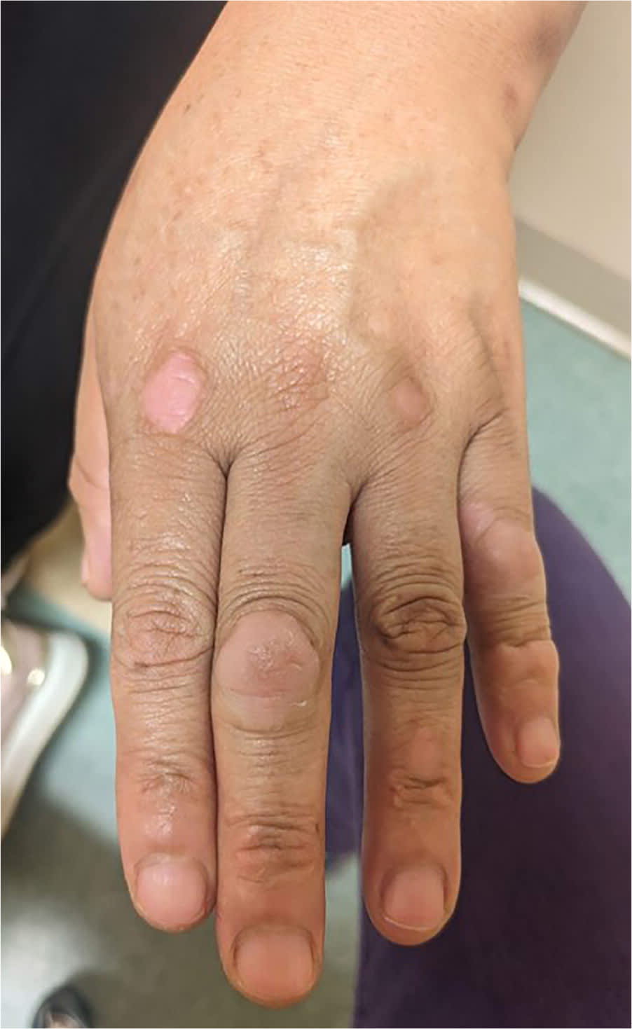
Physical examination of the skin revealed raised, hypopigmented, nontender nodules on the proximal interphalangeal and metacarpophalangeal joints of her left hand (Figure 1). The lesions were slightly pink but had no associated erythema.
FIGURE 1
QUESTION
Based on the patient's history and physical examination, which one of the following is the most likely diagnosis?
- A. Gouty tophi.
- B. Knuckle pads.
- C. Nodular osteoarthritis.
- D. Rheumatoid nodules.
- E. Xanthomas.
DISCUSSION
The answer is B: knuckle pads, which are benign fibromas predominantly localized over the small joints of the hands and feet. They manifest as discrete, well-demarcated, firm, and often painless nodules, typically measuring a few millimeters to centimeters in diameter. The nodules are sometimes hypopigmented. The prevalence of knuckle pads is not well established, but they are common in individuals who do repetitive manual activities, including manual laborers, athletes (eg, boxers, surfers), and musicians. Habits such as chewing or sucking of the fingers and repetitive trauma and pressure to the hands are often seen in patients who develop knuckle pads.
Epidemiologically, knuckle pads are slightly more common in male patients. Although the condition can occur at any age, it is typically seen in adults 30 to 60 years of age. Knuckle pads are associated with several fibrotic disorders, including Dupuytren contracture and Peyronie disease. Knuckle pads are also seen in Bart-Pumphrey syndrome, an autosomal dominant disorder characterized by knuckle pads, leukonychia, sensorineural hearing loss, and palmoplantar keratoderma. Despite these associations, the precise etiology of knuckle pads is unclear.1,2
Characteristic clinical findings include asymptomatic, nontender, slow-growing nodules over the dorsal aspect of the proximal interphalangeal or metacarpophalangeal joints of the hands and the metatarsophalangeal joints of the feet. The nodules are firm and may exhibit overlying skin changes such as hyperkeratosis or callus formation in chronic cases. Knuckle pads can occur unilaterally or bilaterally and may vary in number and size. In some cases, they may be associated with pruritus or discomfort, particularly when the hands or feet are subjected to repetitive trauma or pressure.
Management strategies primarily focus on avoiding repetitive trauma to the hands and feet, minimizing exacerbating factors such as chewing or sucking of the fingers, and closely monitoring any changes in the size, number, and associated symptoms of the lesions. Surgical excision may be considered in symptomatic or cosmetically bothersome cases but is usually reserved for refractory or severe presentations due to the risk of recurrence and potential complications.1–3
Read the full article
Get immediate access, anytime, anywhere.
Choose a single article, issue, or full-access subscription.
Earn up to 14 CME credits per issue.
