

Family physicians play a pivotal role in providing timely care for common fingertip injuries and minimizing long-term disability after injury. Subungual hematoma is diagnosed clinically and treated with observation or nail trephination. Nail bed lacerations are treated by removal of the nail and repair of the nail bed. Distal interphalangeal joint dislocations typically occur dorsally and result from hyperextension or hyperflexion during sports or because of accidental trauma. Management primarily involves closed reduction and splinting; however, surgical intervention might be necessary for complex cases. Distal phalanx fractures are most often minimally displaced and amenable to closed reduction and splinting; open and intra-articular fractures involving more than one-third of the articular surface require referral to a hand surgeon. Mallet finger comprises rupture of the extensor digitorum tendon or avulsion from the adjacent distal phalanx, inhibiting distal interphalangeal joint extension, and typically heals after 6 to 8 weeks of constant immobilization. Jersey finger involves similar injuries to the flexor digitorum profundus tendon but necessitates referral for surgical repair to restore function.
The fingertips play critical roles in everyday activities, from fine motor tasks to tactile sensation. Given the frequent exposure of the fingertips to trauma-inducing stimuli, fingertip injuries are remarkably common, varying broadly in severity and complexity 1 (Table 1). Appropriate diagnosis and management allow family physicians to minimize long-term disability and restore full functionality, underscoring the importance of informed and effective treatment approaches.2,3
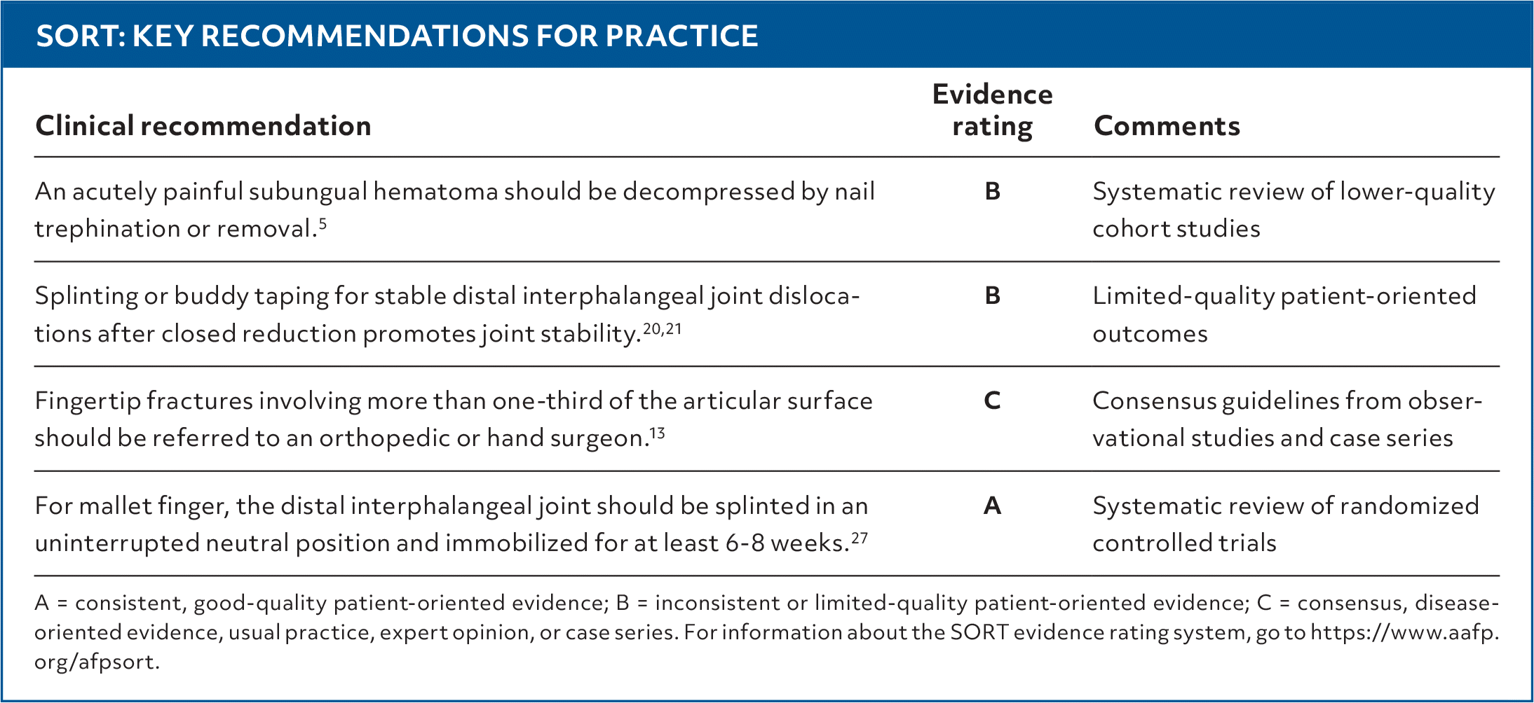
SORT: KEY RECOMMENDATIONS FOR PRACTICE
| Clinical recommendation | Evidence rating | Comments |
|---|---|---|
| An acutely painful subungual hematoma should be decompressed by nail trephination or removal.5 | B | Systematic review of lower-quality cohort studies |
| Splinting or buddy taping for stable distal interphalangeal joint dislocations after closed reduction promotes joint stability.20,21 | B | Limited-quality patient-oriented outcomes |
| Fingertip fractures involving more than one-third of the articular surface should be referred to an orthopedic or hand surgeon.13 | C | Consensus guidelines from observational studies and case series |
| For mallet finger, the distal interphalangeal joint should be splinted in an uninterrupted neutral position and immobilized for at least 6–8 weeks.27 | A | Systematic review of randomized controlled trials |
A = consistent, good-quality patient-oriented evidence; B = inconsistent or limited-quality patient-oriented evidence; C = consensus, disease-oriented evidence, usual practice, expert opinion, or case series. For information about the SORT evidence rating system, go to https://www.aafp.org/afpsort.
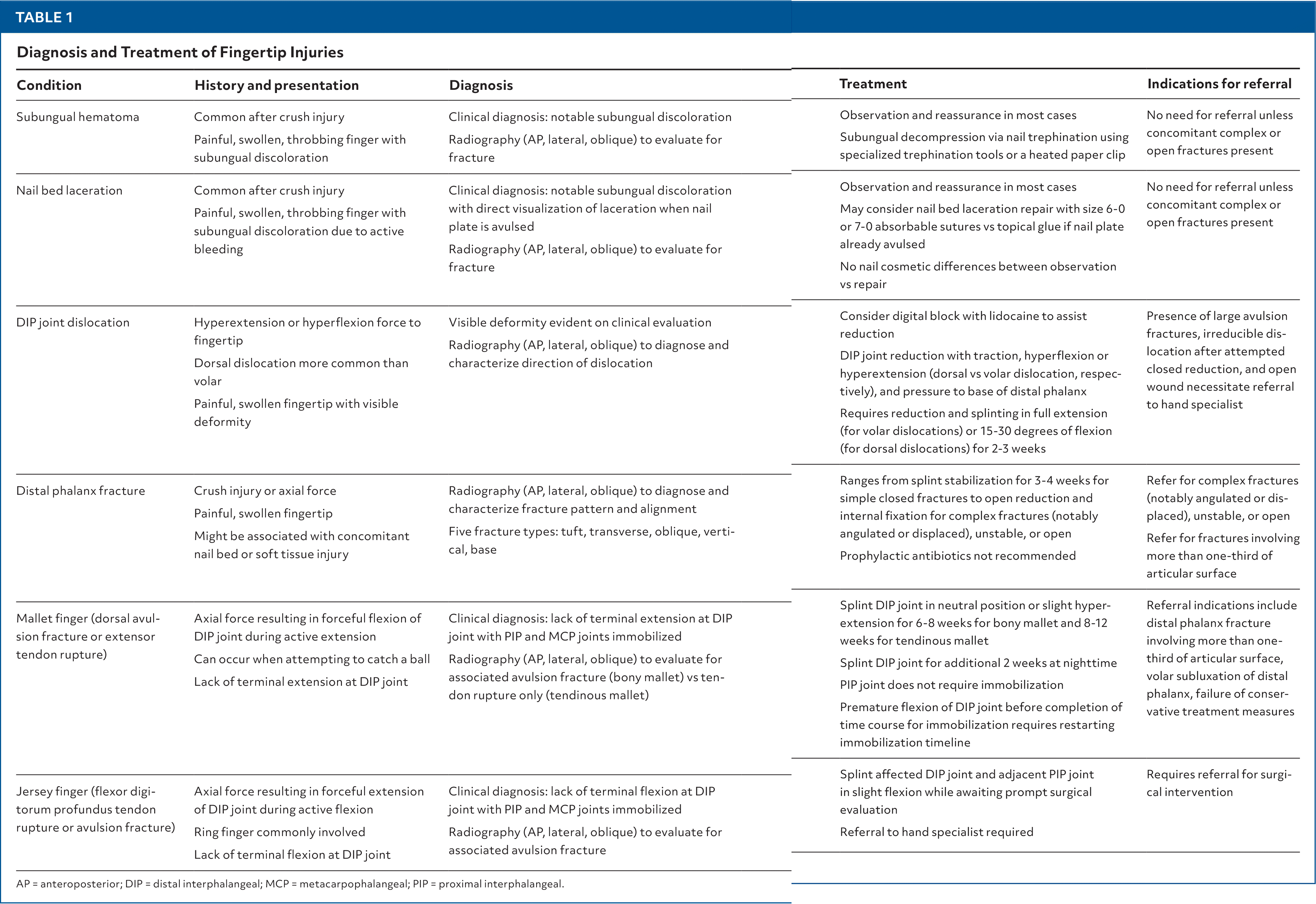
TABLE 1. Diagnosis and Treatment of Fingertip Injuries
| Condition | History and presentation | Diagnosis | Treatment | Indications for referral |
|---|---|---|---|---|
| Subungual hematoma | Common after crush injury Painful, swollen, throbbing finger with subungual discoloration | Clinical diagnosis: notable subungual discoloration Radiography (AP, lateral, oblique) to evaluate for fracture | Observation and reassurance in most cases Subungual decompression via nail trephination using specialized trephination tools or a heated paper clip | No need for referral unless concomitant complex or open fractures present |
| Nail bed laceration | Common after crush injury Painful, swollen, throbbing finger with subungual discoloration due to active bleeding | Clinical diagnosis: notable subungual discoloration with direct visualization of laceration when nail plate is avulsed Radiography (AP, lateral, oblique) to evaluate for fracture | Observation and reassurance in most cases May consider nail bed laceration repair with size 6-0 or 7-0 absorbable sutures vs topical glue if nail plate already avulsed No nail cosmetic differences between observation vs repair | No need for referral unless concomitant complex or open fractures present |
| DIP joint dislocation | Hyperextension or hyperflexion force to fingertip Dorsal dislocation more common than volar Painful, swollen fingertip with visible deformity | Visible deformity evident on clinical evaluation Radiography (AP, lateral, oblique) to diagnose and characterize direction of dislocation | Consider digital block with lidocaine to assist reduction DIP joint reduction with traction, hyperflexion or hyperextension (dorsal vs volar dislocation, respectively), and pressure to base of distal phalanx Requires reduction and splinting in full extension (for volar dislocations) or 15–30 degrees of flexion (for dorsal dislocations) for 2–3 weeks | Presence of large avulsion fractures, irreducible dislocation after attempted closed reduction, and open wound necessitate referral to hand specialist |
| Distal phalanx fracture | Crush injury or axial force Painful, swollen fingertip Might be associated with concomitant nail bed or soft tissue injury | Radiography (AP, lateral, oblique) to diagnose and characterize fracture pattern and alignment Five fracture types: tuft, transverse, oblique, vertical, base | Ranges from splint stabilization for 3–4 weeks for simple closed fractures to open reduction and internal fixation for complex fractures (notably angulated or displaced), unstable, or open Prophylactic antibiotics not recommended | Refer for complex fractures (notably angulated or displaced), unstable, or open Refer for fractures involving more than one-third of articular surface |
| Mallet finger (dorsal avulsion fracture or extensor tendon rupture) | Axial force resulting in forceful flexion of DIP joint during active extension Can occur when attempting to catch a ball Lack of terminal extension at DIP joint | Clinical diagnosis: lack of terminal extension at DIP joint with PIP and MCP joints immobilized Radiography (AP, lateral, oblique) to evaluate for associated avulsion fracture (bony mallet) vs tendon rupture only (tendinous mallet) | Splint DIP joint in neutral position or slight hyper-extension for 6–8 weeks for bony mallet and 8–12 weeks for tendinous mallet Splint DIP joint for additional 2 weeks at nighttime PIP joint does not require immobilization Premature flexion of DIP joint before completion of time course for immobilization requires restarting immobilization timeline | Referral indications include distal phalanx fracture involving more than one-third of articular surface, volar subluxation of distal phalanx, failure of conservative treatment measures |
| Jersey finger (flexor digitorum profundus tendon rupture or avulsion fracture) | Axial force resulting in forceful extension of DIP joint during active flexion Ring finger commonly involved Lack of terminal flexion at DIP joint | Clinical diagnosis: lack of terminal flexion at DIP joint with PIP and MCP joints immobilized Radiography (AP, lateral, oblique) to evaluate for associated avulsion fracture | Splint affected DIP joint and adjacent PIP joint in slight flexion while awaiting prompt surgical evaluation Referral to hand specialist required | Requires referral for surgical intervention |
AP = anteroposterior; DIP = distal interphalangeal; MCP = metacarpophalangeal; PIP = proximal interphalangeal.
Read the full article
Get immediate access, anytime, anywhere.
Choose a single article, issue, or full-access subscription.
Earn up to 14 CME credits per issue.
