

A 12-year-old premenarchal girl presented after 2 days of abdominal pain. She had been seen several times over the previous 6 months for similar symptoms. Her pain had become constant, severe, and sharp, and nonprescription analgesics no longer provided relief. The pain was predominantly in her periumbilical region and radiated to the left lower quadrant. She reported one episode of diarrhea but no nausea, vomiting, anorexia, or fevers. She had no history of recent travel, contact with sick people, dietary changes, or new psychosocial stressors. A review of systems was positive for intermittent constipation and occasional dysuria. She was not sexually active.
Physical examination revealed a tender, palpable mass protruding against her abdominal wall. Computed tomography of the abdomen and pelvis identified the mass as the uterus and vagina, which were both markedly distended with fluid (Figure 1). Magnetic resonance imaging confirmed single uterine and vaginal cavities without abnormal divisions. The cervix and ovaries appeared normal (Figure 2 and Figure 3).
FIGURE 1
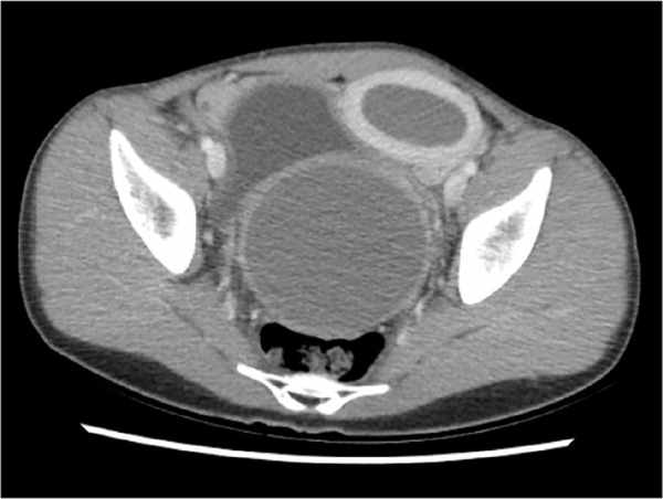
FIGURE 2
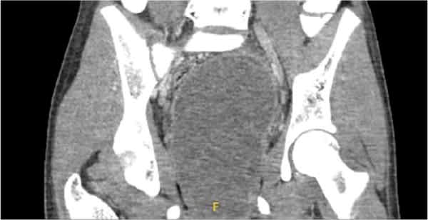
FIGURE 3
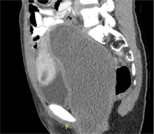
QUESTION
Based on the patient’s history and physical examination, which one of the following is the most likely diagnosis?
- A. Imperforate hymen.
- B. Transverse vaginal septum.
- C. Uterine agenesis.
- D. Uterus didelphys.
DISCUSSION
The answer is A: imperforate hymen, a congenital abnormality in which the hymen does not canalize during embryonic development.1,2 The prevalence is about 1 in 2,000 female patients.2,3
Once menarche occurs, the uterine lining goes through the typical cycle of growth and shedding, but external flow is prevented by the imperforate hymen. This causes the vagina and uterus to become distended with menstrual fluid (a condition known as hematometrocolpos).4 On computed tomography, this patient’s hematometrocolpos appeared as a large homogenous structure in the pelvis that extended into the abdomen.
Hematometrocolpos can cause severe pain, constipation, and urinary issues. When appropriately treated with hymenectomy and evacuation, patients should have no long-term complications or loss of fertility.2,3
A transverse vaginal septum is a congenital abnormality caused by incomplete canalization of the urogenital sinus and müllerian ducts.1,5 The septum can form at various levels within the vagina. Hematometrocolpos may be present. Computed tomography or magnetic resonance imaging shows what appear to be multiple vaginal cavities.
Uterine agenesis occurs when the müllerian system does not develop embryologically, which results in a spectrum of anatomic outcomes.1 A strong genetic predisposition has been identified, and this condition is often seen in family groups.1,6 Anatomic defects outside the genital tract are common. Up to 50% of affected patients have urologic anomalies, and 10% to 15% have issues with skeletal development.6
Uterus didelphys is an anatomic duplication of female reproductive organs, usually the uterus and cervix.1 Amenorrhea is uncommon with this condition.6 Most patients experience few symptoms because the duplicate organs tend to function properly. Uterus didelphys is often diagnosed later in life during evaluation for fertility issues such as recurrent miscarriages.7
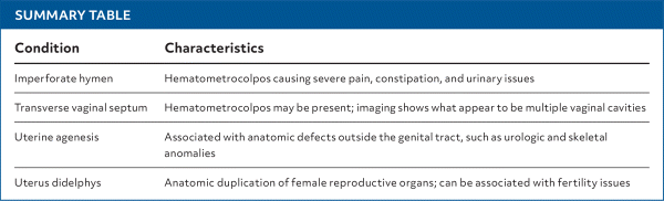
SUMMARY TABLE
| Condition | Characteristics |
|---|---|
| Imperforate hymen | Hematometrocolpos causing severe pain, constipation, and urinary issues |
| Transverse vaginal septum | Hematometrocolpos may be present; imaging shows what appear to be multiple vaginal cavities |
| Uterine agenesis | Associated with anatomic defects outside the genital tract, such as urologic and skeletal anomalies |
| Uterus didelphys | Anatomic duplication of female reproductive organs; can be associated with fertility issues |
Read the full article
Get immediate access, anytime, anywhere.
Choose a single article, issue, or full-access subscription.
Earn up to 14 CME credits per issue.
