

A 5-year-old girl presented with a single small lesion on her back. The lesion had been present for at least 4 years and had not changed significantly in size. It did not bother the patient. She had a history of eczema.
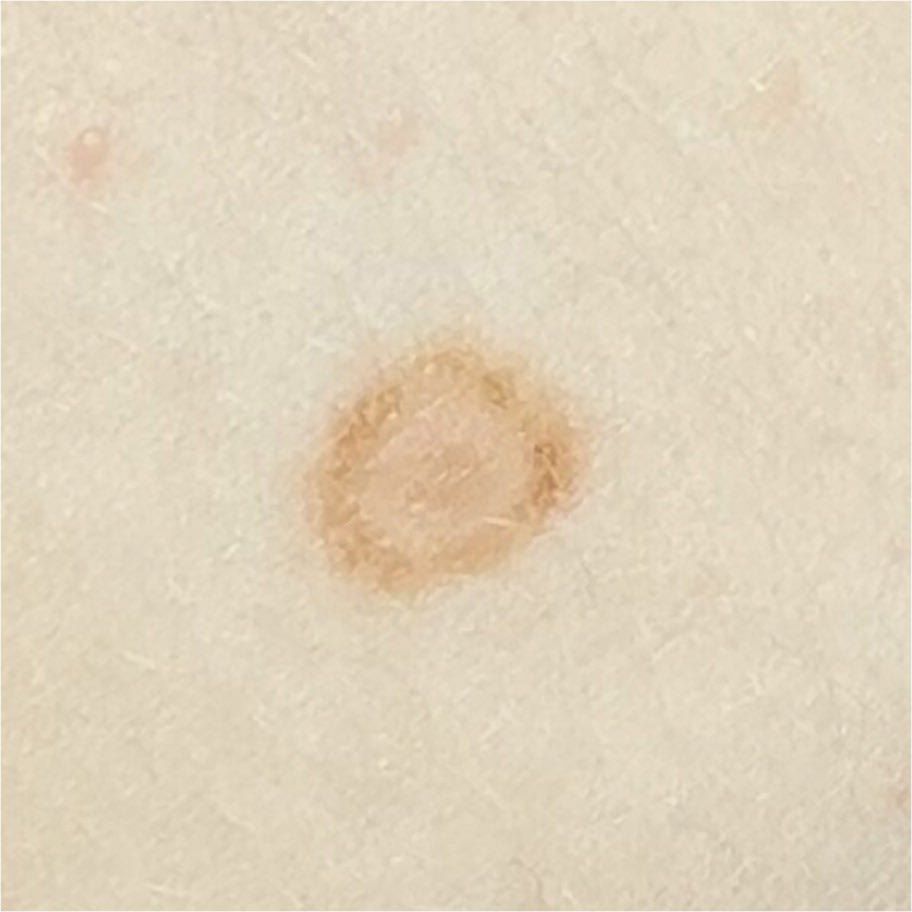
Physical examination revealed a brown, targetoid, macular lesion approximately 6 mm in diameter on the patient's upper back (Figure 1). It was not tender or pruritic.
FIGURE 1
QUESTION
Based on the patient's history and physical examination, which one of the following is the most likely diagnosis?
- A. Cockade nevus.
- B. Eczematous nevus.
- C. Erythema multiforme.
- D. Halo nevus.
DISCUSSION
The answer is A: cockade (or “target”) nevus, a benign lesion that usually occurs in young children on the trunk or scalp. It presents as a central pigmented area encircled by a separate pigmented ring, thus resembling a target.1 On dermoscopic examination, the central pink to brown area is typically homogenous, and the outer pigmented circle appears as a reticular ring.2
Because this is a benign nevus, no further investigation or treatment is necessary.2 However, children with these nevi have a propensity for developing other nevi, which increases the overall risk for melanoma. Parents should be educated on how to perform mole and skin checks.3 Patients with numerous or atypical nevi or a first-degree relative with melanoma should be referred to a dermatologist for regular skin checks.3
An eczematous nevus, also known as a Meyerson nevus, typically presents as a symmetrical area of erythema and scale surrounding a melanocytic nevus. The nevus will persist, but the eczema typically resolves spontaneously within a few months.1
Erythema multiforme lesions classically begin as multiple pink or red papules that progress into “target” or “iris” lesions consisting of a dark center and light pink ring surrounded by a darker red ring. These lesions usually appear in patients younger than 40 years and are often associated with a triggering illness or medication. The lesions resolve in 10 days to 5 weeks.4
A halo nevus, also known as a Sutton nevus, is a benign melanocytic lesion surrounded by an area of hypopigmentation. The average age of onset is 15 years, but this nevus can persist for many years. Most patients have multiple lesions, usually on the back.2
SUMMARY TABLE
| Condition | Characteristics |
|---|---|
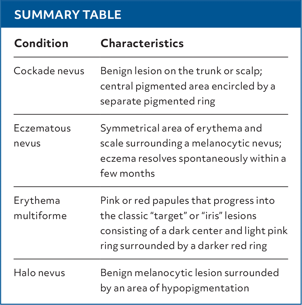
| Cockade nevus | Benign lesion on the trunk or scalp; central pigmented area encircled by a separate pigmented ring |
| Eczematous nevus | Symmetrical area of erythema and scale surrounding a melanocytic nevus; eczema resolves spontaneously within a few months |
| Erythema multiforme | Pink or red papules that progress into the classic “target” or “iris” lesions consisting of a dark center and light pink ring surrounded by a darker red ring |
| Halo nevus | Benign melanocytic lesion surrounded by an area of hypopigmentation |
Read the full article
Get immediate access, anytime, anywhere.
Choose a single article, issue, or full-access subscription.
Earn up to 14 CME credits per issue.
