

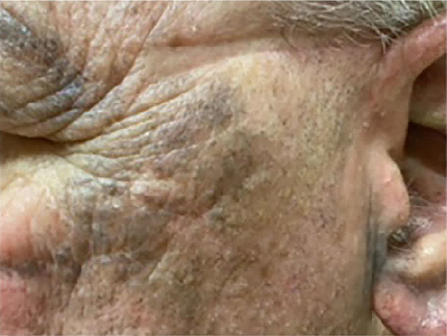
A 71-year-old man presented with progressive black discoloration on the dorsal surfaces of both feet that had been present for 2 years. The discoloration was not associated with pain, burning, or itching. In the previous 6 months, the discoloration had advanced to his distal legs and then separately developed in the preauricular area.
The patient had a history of coronary artery disease, type 2 diabetes, and chronic nodulocystic acne. He was a nonsmoker with no known drug allergies. He had no family history of similar skin findings.
Physical examination confirmed a bluish papular discoloration on both feet as well as his face and ears (Figure 1 and Figure 2). These areas were not tender.
FIGURE 1
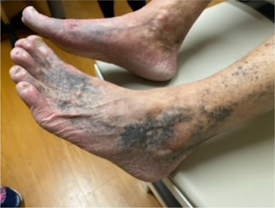
FIGURE 2
QUESTION
Based on the patient's history and physical examination, which one of the following is the most likely diagnosis?
- A. Acanthosis nigricans.
- B. Addison disease.
- C. Chronic venous stasis.
- D. Drug-induced pigmentation.
- E. Melasma.
DISCUSSION
The correct answer is D: drug-induced pigmentation due to long-term minocycline use for treatment of chronic nodulocystic acne. The hyperpigmentation started on the dorsal aspect of the feet with a classic blue and black discoloration. With continued minocycline use, areas of pigmentation darkened and spread proximally.
Hyperpigmentation can appear after long-term ingestion of certain drugs, such as minocycline, usually after years of use or a cumulative dose greater than 50 g. The distribution is variable and can include extensor surfaces of the legs, ankles, dorsa of feet, and face (especially around the eyes), or in sites of inflammation. Pigment deposition has also been reported on internal locations such as bones, cartilage, and the thyroid gland. Discoloration usually disappears gradually over months or years after discontinuation of minocycline; however, pigmentation changes can persist even after discontinuation.1,2
Acanthosis nigricans most commonly appears as a gradual darkening with a velvety texture in skinfold areas such as axillae, neck, groin, or antecubital fossa. It is often associated with diabetes or other insulin-resistant states.3
Addison disease is caused by adrenal insufficiency with loss of cortisol or aldosterone production. [corrected] This results in an overproduction of several pituitary hormones, including melanocyte-stimulating hormone, which then leads to diffuse hyperpigmentation with accentuation in sun-exposed areas and flexural folds.2
Pigmentation from chronic venous stasis is multifactorial. Venous dilation from prolonged venous hypertension increases wall permeability and allows the passage of red blood cells into the interstitium. The destruction of these red cells results in chronic inflammatory changes with fibrin deposition and the release of hemoglobin, which breaks down into hemosiderin. This results in dark brown hyperpigmentation of the distal lower extremities.3,4
Melasma is a macular hyperpigmentation usually found on the upper two-thirds of the face, such as the cheeks, forehead, nose, or upper lip. It can also present on the mandibular surfaces of the face and rarely on the dorsa of the forearms.3
SUMMARY TABLE
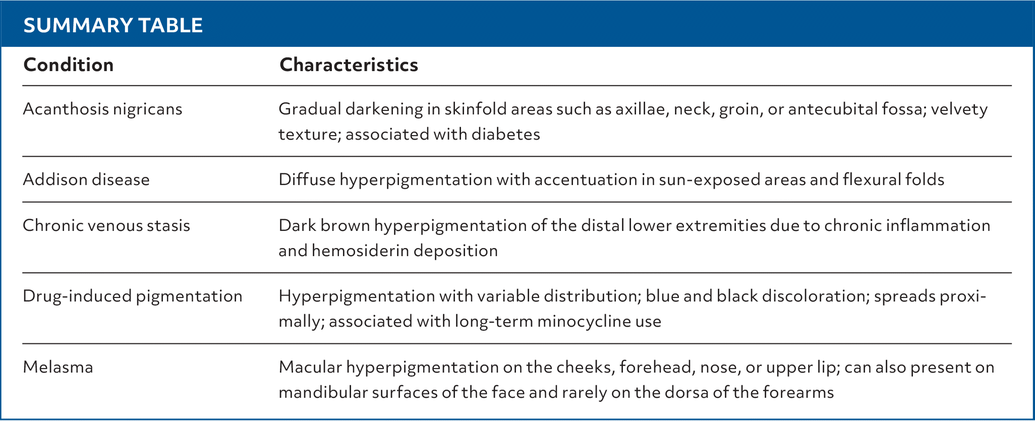
| Condition | Characteristics |
|---|---|
| Acanthosis nigricans | Gradual darkening in skinfold areas such as axillae, neck, groin, or antecubital fossa; velvety texture; associated with diabetes |
| Addison disease | Diffuse hyperpigmentation with accentuation in sun-exposed areas and flexural folds |
| Chronic venous stasis | Dark brown hyperpigmentation of the distal lower extremities due to chronic inflammation and hemosiderin deposition |
| Drug-induced pigmentation | Hyperpigmentation with variable distribution; blue and black discoloration; spreads proximally; associated with long-term minocycline use |
| Melasma | Macular hyperpigmentation on the cheeks, forehead, nose, or upper lip; can also present on mandibular surfaces of the face and rarely on the dorsa of the forearms |
Read the full article
Get immediate access, anytime, anywhere.
Choose a single article, issue, or full-access subscription.
Earn up to 14 CME credits per issue.
