

A 58-year-old man presented with a pruritic rash on his left distal dorsal forearm that had been present for 3 months. It started after his wrist was exposed to paint. The patient had been treated with mupirocin ointment, ketoconazole cream, and desonide 0.05% cream (Desowen). After 3 months of unsuccessful treatment, clobetasol cream had been prescribed, and the rash worsened 1 week later. The patient had a history of untreated pedal onychomycosis and was immunocompetent.
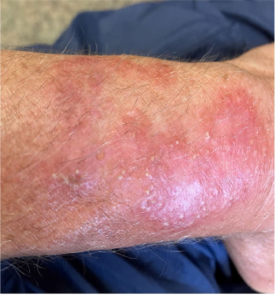
Examination revealed discrete papules, pustules, and nodules coalescing into plaques. The rash was warm and erythematous, with tender scaling plaques and very fine pustules (Figure 1). Gram stain and culture of the pustules showed no bacteria nor growth.
FIGURE 1
QUESTION
Based on the patient's history and physical examination, which one of the following is the most likely diagnosis?
- A. Cellulitis.
- B. Impetiginized contact dermatitis.
- C. Majocchi granuloma.
- D. Methicillin-resistant Staphylococcus aureus folliculitis.
- E. Sporotrichosis.
DISCUSSION
The answer is C: Majocchi granuloma, a rare fungal infection of the dermis. It is commonly caused by dermatophytes, especially Trichophyton rubrum.1,2 Predisposing factors such as tinea infection, trauma, and steroid use were present in this case. Although clinical findings vary in immunocompetent patients, the most common presentations include pruritic, perifollicular, pink-red papules and nodules on the extremities. Other findings include plaques that are warm, red, and painful. The diagnosis is confirmed by biopsy showing granulomatous inflammation around the follicle. Potassium hydroxide testing and fungal culture are also often used for diagnosis, but they do not distinguish between deep and superficial infection. The median time to diagnosis is 44 days.1
The most common treatment for Majocchi granuloma is terbinafine for an average of 31 days, but this may need adjustment based on clinical findings. After shared patient decision-making with a dermatologist, a diagnosis was made clinically. Oral terbinafine and daily ketoconazole cream were started with follow-up after 2 weeks, when a biopsy would be performed if there was no improvement. The pain and warmth improved each day, so a biopsy was not performed. The rash abated after 6 weeks, and 1 year after treatment it had not returned.
Cellulitis presents with warm, red, swollen skin, usually in areas of trauma or injury. It may also present in a postoperative area. Cellulitis is typically not raised or scaling. It is commonly caused by Staphylococcus and Streptococcus species.3
Impetiginized contact dermatitis presents with blistering, pruritus, tenderness, cracking, and swelling. The fine pustules and lack of blisters in this case make impetiginized contact dermatitis unlikely. It can be treated with topical antibiotics but may require oral treatment.
Methicillin-resistant S aureus folliculitis typically presents with more isolated pustules that are larger and more commonly located on the buttock and thigh areas. Treatment requires antibiotics such as clindamycin and doxycycline.4
Sporotrichosis is a fungal infection caused by Sporothrix schenckii. It starts as a single painless, red, purple, or pink papule, and the number of papules increases as the disease progresses. In this case, there was no history of plant or soil exposure, and the coalescing plaques were not consistent with sporotrichosis.5
Read the full article
Get immediate access, anytime, anywhere.
Choose a single article, issue, or full-access subscription.
Earn up to 14 CME credits per issue.
