

A 60-year-old man presented with a rash on his right hand that developed 3 months earlier. The rash was not pruritic, and the patient had no other symptoms or concerns. He reported frequently getting scratches while cleaning his fish tank, predominantly on the affected hand.
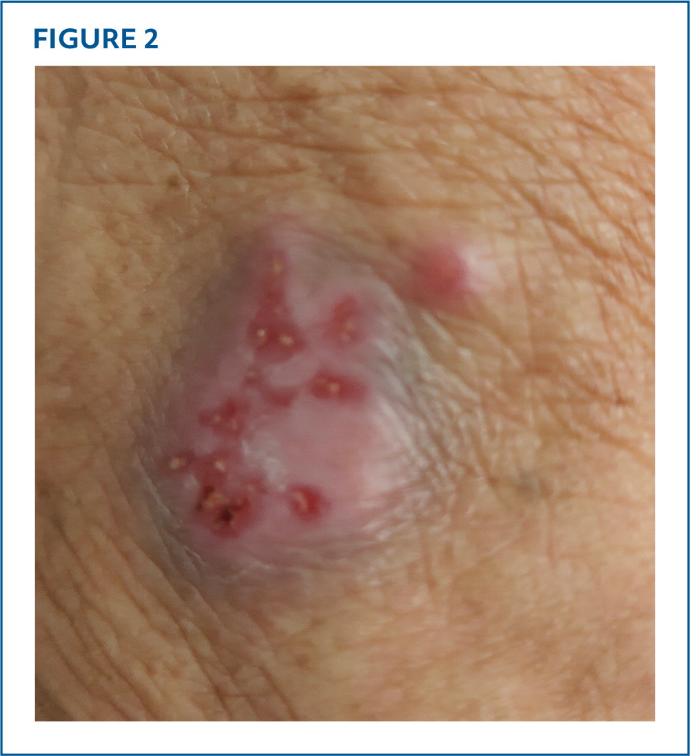
Physical examination revealed a nontender, erythematous rash on the proximal extensor aspect of his right index finger and the area over the metacarpophalangeal joint (Figure 1 and Figure 2). The rash included violaceous papules and signs of scaling. No other skin findings were present. Laboratory testing included a C-reactive protein level of .7 mg/L and white blood cell count of 5,300/μL (5.3 × 10⁹/L). Radiography of the affected hand showed no acute fracture, bony erosion, or periosteal reaction.
FIGURE 1
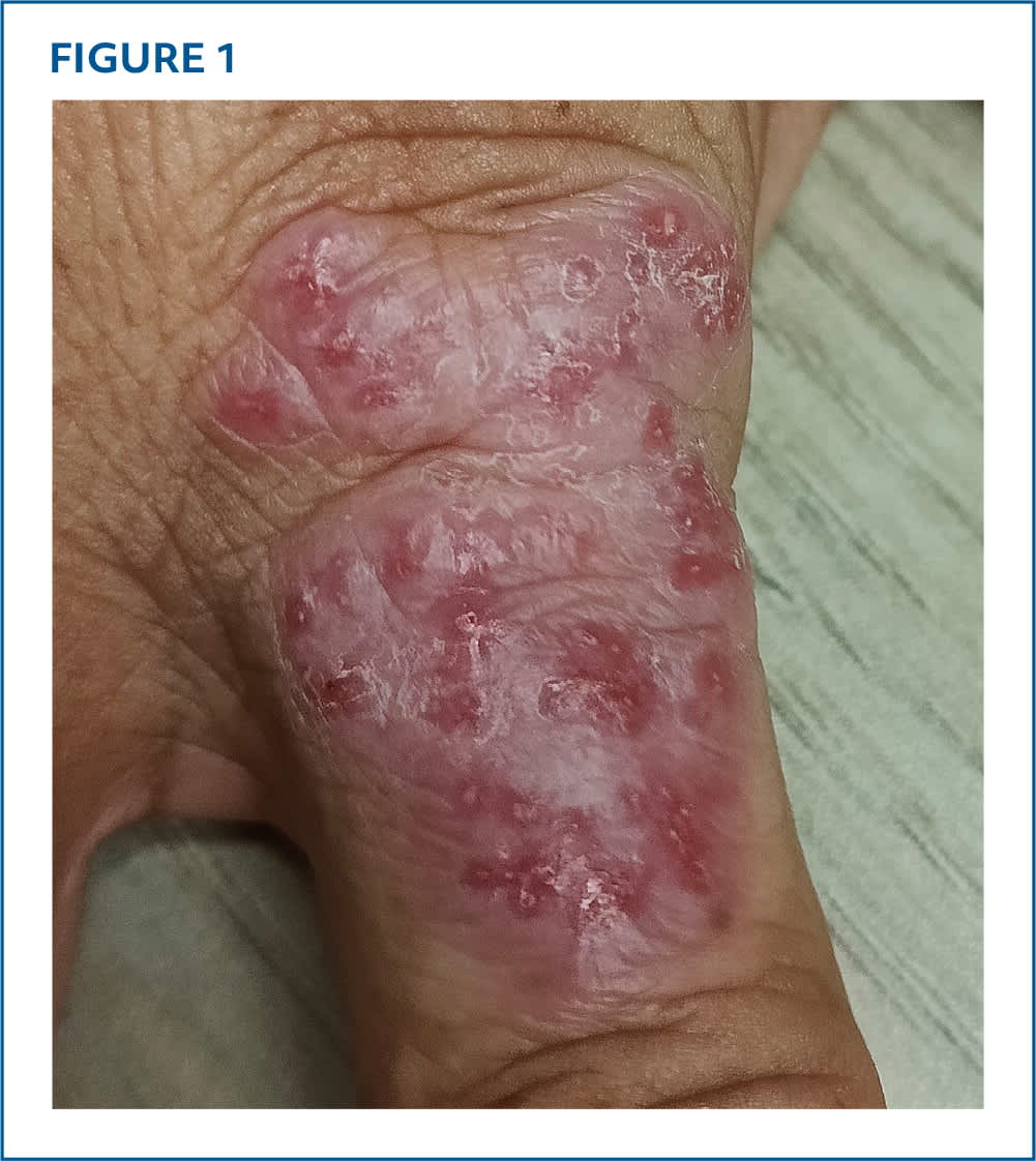
FIGURE 2
QUESTION
Based on the patient's history and physical examination, which one of the following is the most likely diagnosis?
- A. Cutaneous leishmaniasis.
- B. Cutaneous squamous cell carcinoma.
- C. Melioidosis.
- D. Mycobacterium marinum infection.
- E. Sarcoidosis.
DISCUSSION
The answer is D: Mycobacterium marinum infection, also known as fish tank granuloma. In the United States, the prevalence of cutaneous nontuberculous Mycobacterium infection is approximately 1.3 per 100,000 people.1 Although M marinum infection is rare, it constitutes most cases of nontuberculous Mycobacterium infections.2 Infection occurs with continual exposure to contaminated water or direct contact with fish or shellfish. Exposure to swimming pools that are not adequately sanitized may also cause infection.3 The incubation period is 2 to 3 weeks.2
M marinum infection typically presents as a single papulonodular, verrucous, or ulcerated granular lesion at sites of trauma. When multiple lesions develop, the rash can form a sporotrichoid pattern on exposed skin, typically on the extremities. The rash usually develops 2 to 8 weeks after bacterial inoculation. M marinum infection is diagnosed clinically but can be confirmed histologically with skin biopsy. Treatment requires two active anti-mycobacterial agents, usually macrolides plus rifampin, with guidance from susceptibility tests.3
Cutaneous leishmaniasis typically presents a few weeks or months after a sand fly bite. It causes a painless papular or nodular rash that can progress into ulcers with hard, raised borders and sunken centers.4
Cutaneous squamous cell carcinoma can present as a gradually enlarging ulcer with raised edges, or an asymptomatic ulcerated nodule or plaque. Lesions typically occur at sites of chronic exposure, such as on the face and dorsa of hands.5
Melioidosis is a bacterial infection caused by Burkholderia pseudomallei, which is found in contaminated soil and water, particularly in tropical climates such as Southeast Asia and northern Australia. Melioidosis usually presents with a solitary, chronic, nonhealing ulcer that does not respond well to antibiotics, often on the lower legs. Patient may also present with lymphadenopathy and constitutional symptoms such as fever and lethargy.6,7
Sarcoidosis may cause erythema nodosum, typically on the shins. The nodules are tender and dark reddish-brown to erythematous. Sarcoidosis may resolve spontaneously.8
SUMMARY TABLE
| Condition | Characteristics |
|---|---|
| Cutaneous leishmaniasis | Painless papular or nodular rash that can ulcerate |
| Cutaneous squamous cell carcinoma | Gradually enlarging lesion with raised edges, or an asymptomatic ulcerated nodule or plaque |
| Melioidosis | Solitary, chronic, nonhealing ulcer |
| Mycobacterium marinum infection | Papulonodular, verrucous, or ulcerated granular lesion at sites of trauma in a sporotrichoid pattern |
| Sarcoidosis | Tender, dark reddish-brown to erythematous nodules |
Read the full article
Get immediate access, anytime, anywhere.
Choose a single article, issue, or full-access subscription.
Earn up to 14 CME credits per issue.
