

A 30-year-old woman with type 2 diabetes and hypertension presented with an asymptomatic, slowly enlarging growth on the anterior aspect of her right hip. The lesion had been present for at least 1 year. She did not have any other lesions but reported that several family members had similar growths removed.
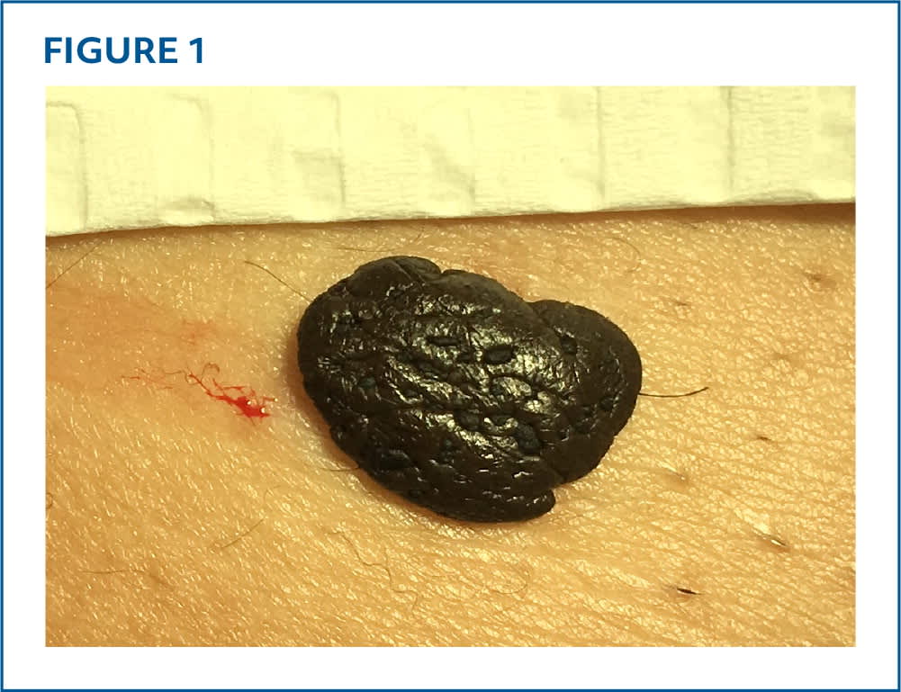
Physical examination revealed a dark brown, verrucous, hyperpigmented plaque that was 10 × 13 × 5 mm in size (Figure 1). The remainder of the skin examination was unremarkable.
FIGURE 1
QUESTION
Based on the patient's history and physical examination, which one of the following is the most likely diagnosis?
- A. Dysplastic nevus.
- B. Melanocytic nevus.
- C. Nodular melanoma.
- D. Pigmented basal cell carcinoma.
- E. Seborrheic keratosis (cutaneous melanoacanthoma type).
DISCUSSION
The answer is E: seborrheic keratosis (cutaneous melanoacanthoma type). Seborrheic keratoses are common benign skin tumors. They are usually sharply demarcated and can vary in color, size, and surface characteristics (eg, flat, smooth, verrucous, scaly, waxy). They are often flat, raised, or pedunculated, giving the classic “stuck-on” appearance.1
Some argue that cutaneous melanoacanthoma is a clinically and histologically distinct entity based on the distribution of dendritic melanocytes, but others consider it a rare variant of seborrheic keratosis notable for its deep pigmentation.2 Cutaneous melanoacanthoma presents as a single, slow-growing, deeply pigmented plaque or nodule with a median diameter of 2 cm. Some lesions may feature multiple colors. Cutaneous melanoacanthoma is more common in men, with a median age of 65 years. Lesions typically occur on the head, neck, trunk, or extremities. Although the exact pathogenesis has not yet been fully established, reactivity related to local irritation or trauma has been implicated.2
Seborrheic keratoses are often removed for cosmetic reasons. They can become inflamed by unconscious manipulation, chafing from clothing, or maceration in intertriginous areas.1 If the diagnosis is certain, no treatment is necessary.1 The most clinically significant condition to exclude is malignant melanoma. Biopsy should be performed for histologic examination to confirm the diagnosis if necessary.1
Dysplastic nevi (ie, atypical moles) result from a benign proliferation of melanocytes. Malignant progression is possible but rare.3,4 Dysplastic nevi typically appear during puberty but can develop at any age. They can present in multiple forms, occurring most often on the trunk and extremities. Dysplastic nevi are typically more than 5 mm in diameter and asymmetrical, with irregular borders and variable color distributions.3,4
Melanocytic nevi also result from a benign proliferation of melanocytes. Predisposing factors include genetic disposition, sun exposure, fair skin tone, and immunosuppression. Lesions typically appear after 6 months of age, increase in number during childhood, and resolve in adulthood. They are usually small (less than 6 mm in diameter), symmetrical, and round or oval with uniform pigmentation and a sharply demarcated border. No treatment is necessary, but lesions may be removed for cosmetic reasons.5,6
Nodular melanoma resembles other benign lesions such as dermal nevi, seborrheic keratosis, or dermatofibroma and does not conform to the usual ABCD (asymmetry, border irregularity, color variation, diameter of 6 mm or more) criteria for melanoma diagnosis.7 This rapidly growing malignancy is more common in males than females (with a ratio of 2: 1), usually occurs in patients 40 to 50 years of age, and can arise anywhere on the body. It presents as an asymmetrical, unevenly pigmented, elevated, firm, pedunculated or polypoid nodule that may become ulcerated in later stages.7
Read the full article
Get immediate access, anytime, anywhere.
Choose a single article, issue, or full-access subscription.
Earn up to 14 CME credits per issue.
