

A 60-year-old man with a history of basal and squamous cell carcinoma of the skin presented for follow-up. He had no skin-related concerns.
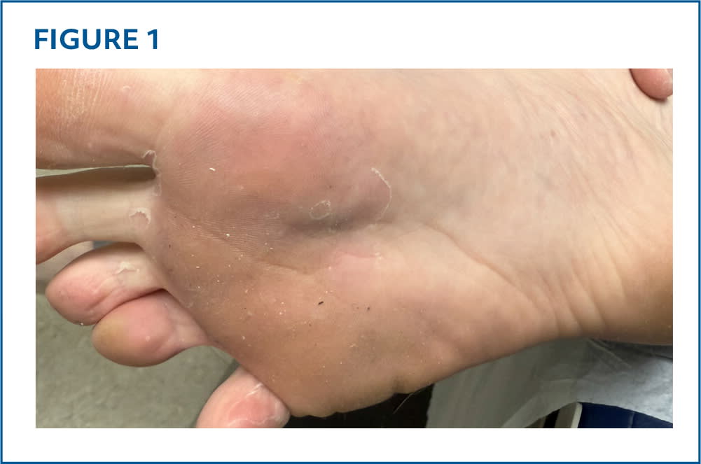
Physical examination revealed circular, smooth macules and small patches with peripheral collarettes of desquamative scales on both soles (Figure 1) that had been present for many years. Warm temperatures and sweating made the peeling worse. He had no associated symptoms such as itching or pain. His nails were normal.
FIGURE 1
QUESTION
Based on the patient's history and physical examination, which one of the following is the most likely diagnosis?
- A. Acral peeling skin syndrome.
- B. Atopic dermatitis.
- C. Dyshidrotic eczema.
- D. Keratolysis exfoliativa.
- E. Tinea pedis.
DISCUSSION
The answer is D: keratolysis exfoliativa, a superficial desquamative palmoplantar dermatosis that is more common on the palms than the soles. The typical lesion begins as an annular pink macule with a central air-filled blister, which later ruptures leaving a collarette of scales, as seen in this patient. More severe cases may present with multiple coalescing patches that form large areas of desquamation.1 The keratolysis exfoliative rash is usually asymptomatic, but burning or pruritus may occur.1,2 Keratolysis exfoliativa may be under- or misdiagnosed and is not frequently reported in the literature.2
The pathophysiology of keratolysis exfoliativa is unclear, but there is some evidence that premature corneodesmolysis in the stratum corneum may play a role. Friction, water, and warm environments may worsen keratolysis exfoliativa. Some studies support an association with palmoplantar hyperhidrosis.1
Education is the cornerstone of treatment. Patients should be instructed to limit exposure of the palms and soles to moisture, harsh cleansers, hot environments, and chronic friction. Keratolytic agents containing urea, ammonium lactate, or salicylic acid can help exfoliate the desquamative scale.3 Given the lack of underlying inflammation, keratolysis exfoliativa does not typically respond to topical corticosteroids or calcineurin inhibitors.4 The condition is usually asymptomatic and can be an incidental finding; therefore, treatment is often not needed.
Acral peeling skin syndrome is a rare dermatosis caused by a TGM5 or CSTA gene variation.4 It usually presents soon after birth with superficial peeling or air-filled blisters of the dorsal and palmoplantar aspects of the hands and feet, although it can also affect the arms and legs.1
Atopic dermatitis is a chronic inflammatory dermatitis that can occur at any age. It most commonly affects flexural surfaces, specifically the back of the neck and the antecubital and popliteal fossae. Dorsal, and less commonly palmoplantar, surfaces of the hands, feet, legs, and arms can be involved. Pink scaly patches, often with lichenification and excoriations, are common. Extreme pruritus is the hallmark of this condition.2,5
Dyshidrotic eczema is a chronic dermatosis characterized by small pruritic vesicles filled with clear fluid. They typically occur on the lateral aspect of the digits but can also involve the palms and soles.1
Tinea is a common fungal infection that can affect hair, skin, and nails. Involvement of the feet (tinea pedis) typically occurs between the toes but can also present as diffuse hyperkeratotic scaly patches on the soles, often in a moccasin distribution. Collarettes of scale can be found on the lateral aspect of the feet but without antecedent air-filled blisters. Involvement of both soles and one palm, or one sole and both palms can occur.6 Thickening and yellowing of the toenail plates are common, often with subungual hyperkeratosis. Potassium hydroxide microscopic examination of a scale scraping will demonstrate branching hyphae.1
Read the full article
Get immediate access, anytime, anywhere.
Choose a single article, issue, or full-access subscription.
Earn up to 14 CME credits per issue.
