

A 39-year-old otherwise healthy woman presented with a painful, burning rash on her hands and feet that appeared 2 days earlier and progressively worsened. One day before the rash appeared, she developed a sore throat and low-grade fever (100.7°F [38.2°C]). Her 5-month-old child attended day care, but no other household members had similar symptoms.
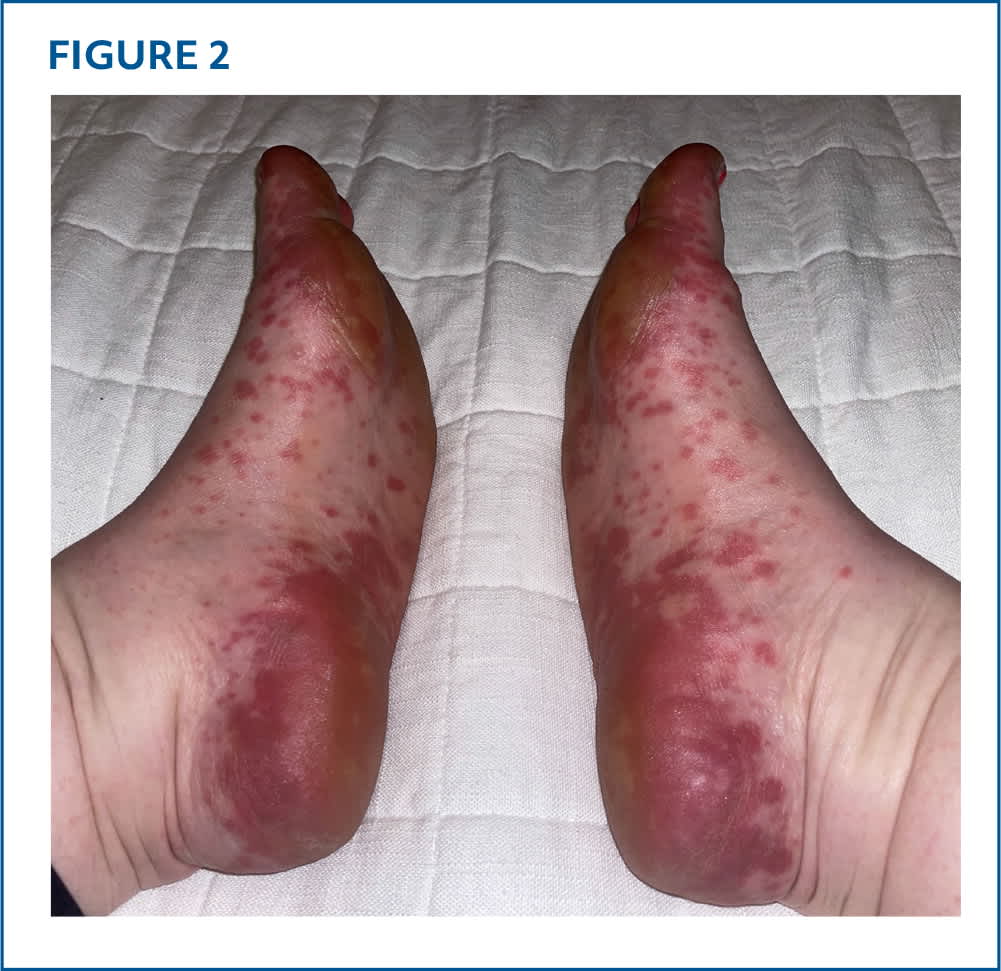
Physical examination showed multiple well-demarcated, erythematous macules diffusely scattered over bilateral palms and soles, interdigital spaces, and lateral and dorsal aspects of the hands and feet (Figures 1 and 2). On some areas of the palms and soles, macules coalesced to form large erythematous patches. Oral examination showed ulcerations on her soft palate. The patient was afebrile at presentation, and the remainder of her physical examination was unremarkable.
FIGURE 1
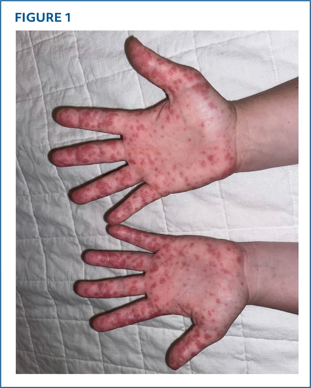
FIGURE 2
QUESTION
Based on the patient's history and physical examination, which one of the following is the most likely diagnosis?
- A. Cutaneous small vessel vasculitis.
- B. Erythema multiforme.
- C. Hand-foot-and-mouth disease.
- D. Reactive infectious mucocutaneous eruption.
- E. Rocky Mountain spotted fever.
DISCUSSION
The answer is C: hand-foot-and-mouth disease, an acute viral illness most commonly caused by coxsackievirus A16 and enterovirus 71.1,2 The disease is highly contagious and may be transmitted through fecal-oral, oral-oral, and respiratory droplet routes.
Hand-foot-and-mouth disease typically presents as a prodrome of low-grade fever, sore throat, and malaise followed by the characteristic maculopapular or vesicular rash on the palms and soles, and occasionally on the buttocks, genitals, knees, and elbows. Painful oral ulcerations occur on the buccal mucosa, tongue, and palate.1
Although hand-foot-and-mouth disease predominantly affects children younger than 10 years, adults can be infected, particularly through household contact with infected children.3 The incubation period is typically 3 to 7 days, and lesions usually resolve within 7 to 10 days.
Coxsackievirus A6 has emerged as a significant pathogen associated with more severe, atypical presentations, including widespread cutaneous involvement, vesiculobullous eruptions, purpuric lesions, and eczema coxsackium (superinfection of eczematous skin). Late sequelae such as onychomadesis (nail shedding) and palmoplantar desquamation may occur 3 to 8 weeks after coxsackievirus A6 infection.2
Treatment of hand-foot-and-mouth disease is supportive, focusing on hydration and pain management with acetaminophen or ibuprofen. Oral lidocaine is not recommended, and no specific antiviral therapy is approved for use. Prevention relies on handwashing and disinfecting contaminated surfaces.1
Cutaneous small vessel vasculitis can develop 7 to 10 days after a provoking drug or infection. Presentation includes palpable purpura (0.3–1 cm in diameter) and nonblanching petechiae (purpuric lesions less than 3 mm in diameter) predominantly on lower extremities. Lesions may coalesce, ulcerate, and be surrounded by hemorrhagic bullae.4
Erythema multiforme is a hypersensitivity reaction often triggered by herpes simplex virus infection or medications. It is characterized by targetoid lesions with concentric color variation. It classically presents on acral sites and progresses centrally.1 Erythema multiforme major may be associated with mucosal involvement and systemic symptoms such as fever and arthralgias.
Reactive infectious mucocutaneous eruption is a postinfectious condition characterized by prominent mucositis involving two or more sites (oral, ocular, and urogenital) with minimal cutaneous involvement. Reactive infectious mucocutaneous eruption is precipitated by infection with bacteria or viruses such as Mycoplasma pneumoniae or adenovirus.1,5
Read the full article
Get immediate access, anytime, anywhere.
Choose a single article, issue, or full-access subscription.
Earn up to 14 CME credits per issue.
