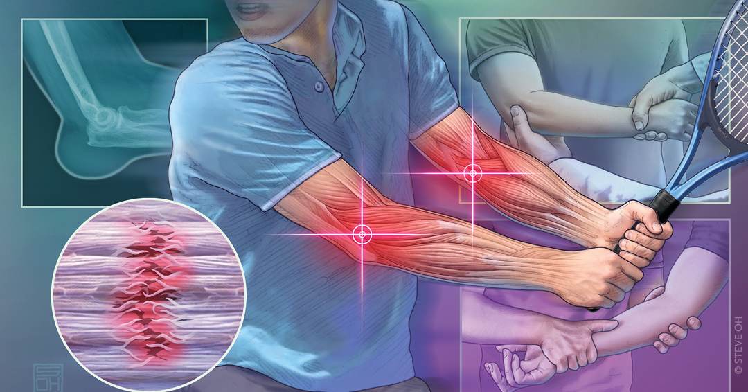
Elbow pain is a common musculoskeletal condition seen in primary care. It most often results from overuse tendinopathies, neuropathic compression, traumatic injuries, or intra-articular disorders. A regional diagnostic approach that localizes pain to the anterior, lateral, medial, or posterior elbow efficiently narrows the differential diagnosis and guides a targeted physical examination. Examination findings, including point tenderness, painful resisted motions, and limitations in passive vs active range of motion, can help distinguish periarticular soft tissue conditions from intra-articular pathology. Red flag features such as high-energy trauma, systemic symptoms, rapid swelling and erythema, or progressive neurologic deficits warrant urgent evaluation. Radiography is appropriate for suspected fracture, deformity, or mechanical symptoms; musculoskeletal ultrasonography can accurately assess tendon and nerve pathology. Magnetic resonance imaging is reserved for cases in which symptoms persist despite conservative care or when advanced structural injury is suspected.
Elbow pain is a common condition seen in primary care. It most often results from overuse tendinopathies, neuropathic compression, traumatic injuries, or intra-articular disorders, including degenerative, inflammatory, and infectious causes. A regional diagnostic approach that localizes pain to the anterior, lateral, medial, or posterior elbow narrows the differential diagnosis and guides a targeted physical examination. Table 1 lists the differential diagnosis of elbow pain in adults.1–24
Lateral elbow tendinopathy affects an estimated 1% to 3% of the general adult population and is one of the most common diagnoses.9 Among specific elbow diagnoses in outpatient cohorts, lateral elbow tendinopathy accounts for more than one-half of cases, followed by medial elbow tendinopathy.9,25,26 Risk of elbow tendinopathy increases with repetitive gripping, wrist motion, and heavy lifting. Elbow tendinopathy is most common in the dominant arm of patients 35 to 55 years of age.15,27–29
Urgent evaluation is warranted for suspected infection, acute dislocation, compartment syndrome, or tendon rupture. Red flags include fever or systemic illness, rapidly progressive swelling or erythema, pain out of proportion to examination findings, inability to move the elbow, neurovascular compromise, progressive neurologic deficits, and high-energy trauma.7,8,24,30 Acute traumatic elbow injuries, including fractures and dislocations, are not included in this review and are addressed in detail in a previous American Family Physician article.31
SORT: KEY RECOMMENDATIONS FOR PRACTICE
| Clinical recommendation | Evidence rating | Comments |
|---|---|---|
| Use the hook test to evaluate for suspected distal biceps tendon rupture and refer urgently when the test is abnormal.1,4 | C | Prospective diagnostic studies that demonstrated high specificity for complete distal biceps rupture |
| Suspect lateral elbow tendinopathy in patients with pain localized at approximately 1 cm distal to the lateral epicondyle that is reproduced with resisted wrist extension or middle-finger extension; grip strength may also be reduced.10 | C | Systematic reviews of diagnostic accuracy studies |
| Suspect ulnar collateral ligament injury in overhead-throwing athletes with medial elbow pain and a positive moving valgus stress test.20 | C | Small diagnostic accuracy study with high sensitivity |
| Reserve bursal aspiration for diagnostic uncertainty, refractory symptoms, or suspected septic olecranon bursitis; routine aspiration of uncomplicated aseptic olecranon bursitis should be avoided because of the risk of complications.24,33,40,42 | C | Randomized and observational data |
| Perform plain radiography as initial imaging for acute elbow trauma, deformity, or mechanical symptoms.43,44 | C | Expert consensus and imaging guidelines |
| Use musculoskeletal ultrasonography to evaluate suspected lateral elbow tendinopathy, olecranon bursitis, or ulnar nerve enlargement in cubital tunnel syndrome when expertise is available.2,45,48 | C | Reviews and expert guidance |
A = consistent, good-quality patient-oriented evidence; B = inconsistent or limited-quality patient-oriented evidence; C = consensus, disease-oriented evidence, usual practice, expert opinion, or case series. For information about the SORT evidence rating system, go to https://www.aafp.org/afpsort.
Read the full article
Get immediate access, anytime, anywhere.
Choose a single article, issue, or full-access subscription.
Earn up to 14 CME credits per issue.