A 68-year-old woman presented to the dermatology clinic with persistent lesions on her trunk and extremities. Two years earlier, she had consulted her primary care physician regarding four scattered “lumps,” which she attributed to mosquito bites. Two years later, she was seen by a different primary care physician when she developed discoloration on her arm, which she believed was related to an aspirin allergy. The patient was seen twice more over the following 3 months before referral to the dermatology clinic.
The patient had a history of breast cancer 30 years previously, which was treated with radical mastectomy, chemotherapy, and radiation. She also had a history of controlled hypertension, type 2 diabetes mellitus, and an allergic reaction to iodine contrast media.
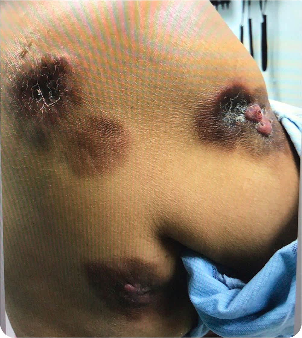
Examination revealed multiple firm, painless, hyperpigmented plaques scattered across her trunk and extremities (Figure 1). The plaques contained central pink nodules.
FIGURE 1
Question
Based on the patient's history and physical examination, which one of the following is the most likely diagnosis?
- A. Angiosarcoma (Stewart-Treves syndrome).
- B. Cutaneous metastasis from breast cancer.
- C. Discoid lupus erythematosus.
- D. Fixed drug eruption.
- E. Keloids.
Discussion
The correct answer is B: cutaneous metastasis from breast cancer. The clinical presentation of cutaneous metastasis is broad and can mimic benign conditions such as keloids, dermatofibromas, prurigo nodularis, and discoid lupus erythematosus. The most common presentation is the combination of plaques and erythematous nodules involving the chest and arms, as in this case.1 The diagnosis depends on the clinical examination, as well as the histopathologic and immunohistochemistry analysis of a biopsy specimen.2 Lesions should be assessed for duration, growth, associated pruritus, bleeding, discharge, and pain. Any lesion demonstrating notable growth, pain, or bleeding requires further evaluation to exclude malignancy. Physicians should also ask the patient whether they have any history of malignancy.
This case illustrates the importance of early biopsy for diagnosis of skin lesions in patients who have had a history of cancer. The possibility of cutaneous metastasis must be considered, even years after treatment of the primary condition.2 Breast cancer is the leading cause of cutaneous metastasis, which is an unusual finding that suggests substantial disease progression.2,3 Patients with the condition are typically 50 to 70 years of age. Symptoms occur relatively soon after the initial cancer diagnosis, often within 3 years.2
Cutaneous metastasis typically presents as multiple firm, nontender nodules that appear suddenly, grow rapidly, and often demonstrate a fixed adherent nature to underlying tissues.4
Angiosarcoma, also known as Stewart-Treves syndrome, is a malignant tumor of the blood or lymph vessels. It can appear as an area of ecchymosis or a violaceous to red papule. The tumor can progress to invasion of underlying tissue, which may cause edema, ulceration, and bleeding due to tumor enlargement.5
Discoid lupus erythematosus classically appears on the face, ears, and scalp. Lesions often consist of erythematous or violaceous macules and scaling plaques. These lesions can lead to scarring, dyspigmentation, and alopecia.4
A fixed drug eruption is a unique form of drug allergy that produces red plaques or blisters that recur at the same cutaneous or mucosal site due to subsequent ingestion of the specific drug.4
Keloids are thickened scar tissue, which may involve the chest and shoulders.4 Patients often have a history of trauma, surgery, insect bites, vaccinations, burns, skin piercings, or acne.6 Keloids will grow beyond the initial border of a scar.4 In the authors' experience, keloids tend to demonstrate less of a fixed, hardened, and bound-down nature to underlying tissues, which may occur with cutaneous metastatic disease.
SUMMARY TABLE
| Condition | Characteristics |
|---|---|
| Angiosarcoma (Stewart-Treves syndrome) | Malignant tumor; can initially appear as an area of ecchymosis or a violaceous to red papule, with progression to invasion of underlying tissue; may cause edema, ulceration, and bleeding due to tumor enlargement |
| Cutaneous metastasis from breast cancer | Personal history of cancer; multiple firm, nontender nodules that appear suddenly, grow rapidly, and often demonstrate a fixed adherent nature to underlying tissues |
| Discoid lupus erythematosus | Often consists of erythematous or violaceous macules and scaling plaques; can lead to scarring, dyspigmentation, and alopecia |
| Fixed drug eruption | Unique form of drug allergy that produces red plaques or blisters; recurs at the same cutaneous or mucosal site due to subsequent ingestion of the specific drug |
| Keloids | Thickened scar tissue that may occur on the chest and shoulders; patients often have a history of trauma, surgery, insect bites, vaccinations, burns, skin piercings, or acne; will grow beyond the initial border of a scar |
The views expressed in this Photo Quiz are those of the authors and do not necessarily reflect the official policy or position of the Department of the Navy, Department of Defense, or the U.S. government.
I am an employee of the U.S. government. This work was prepared as part of my official duties. Title 17 U.S.C. 105 provides that “Copyright protection under this title is not available for any work of the United States Government” Title 17 U.S.C. 101 defines a United States Government work as a work prepared by a military service member or employee of the United States Government as part of that person’s official duties.
