Pregnancy dating is determined by the patient's last menstrual period or an ultrasound measurement. A full-term pregnancy is considered 37 weeks' gestation or more. Spontaneous labor begins when regular painful uterine contractions result in a cervical change. Active labor begins at 6 cm dilation and is marked by more predictable, accelerated cervical change. In the absence of pregnancy complications, intermittent fetal auscultation may be considered as an alternative to continuous electronic fetal monitoring, which is associated with a high false-positive rate. Intravenous antibiotic prophylaxis is indicated in patients with group B streptococcus colonization or those at high risk to prevent newborn early-onset group B streptococcus. The likelihood of vaginal delivery is increased by providing continuous nonmedical support during labor, encouraging mobility, and using a peanut ball with epidural analgesia. Neuraxial analgesia is more effective for pain control than systemic opioids and is associated with fewer adverse effects. Delayed pushing during the second stage of labor has risks but does not affect the mode of delivery. Routine oropharyngeal suctioning of the newborn is not recommended, even with meconium-stained amniotic fluid. Delayed cord clamping reduces newborn anemia. Prevention of postpartum hemorrhage in patients at risk includes prophylactic uterotonic administration and controlled cord traction. Perineal lacerations that alter anatomy or are not hemostatic should be repaired. (Am Fam Physician. 2024;109(6):525-532. Copyright © 2024 American Academy of Family Physicians.)
Approximately 2.5 million infants were born via spontaneous vaginal delivery in the United States in 2021.1 The estimated due date of a pregnancy is 40 weeks after the first day of the patient's last menstrual period; a full-term pregnancy is considered 37 weeks' gestation or more.2 Pregnancy dating can be confirmed by an ultrasound measurement of the fetal crown-rump length early in the first trimester. Ultrasound dating should be used for irregular or unknown menstrual timing or a significant discrepancy between menstrual and ultrasound dating (Table 1).2 Accurate dating guides surveillance and management, including the timing of medically indicated delivery when needed.3
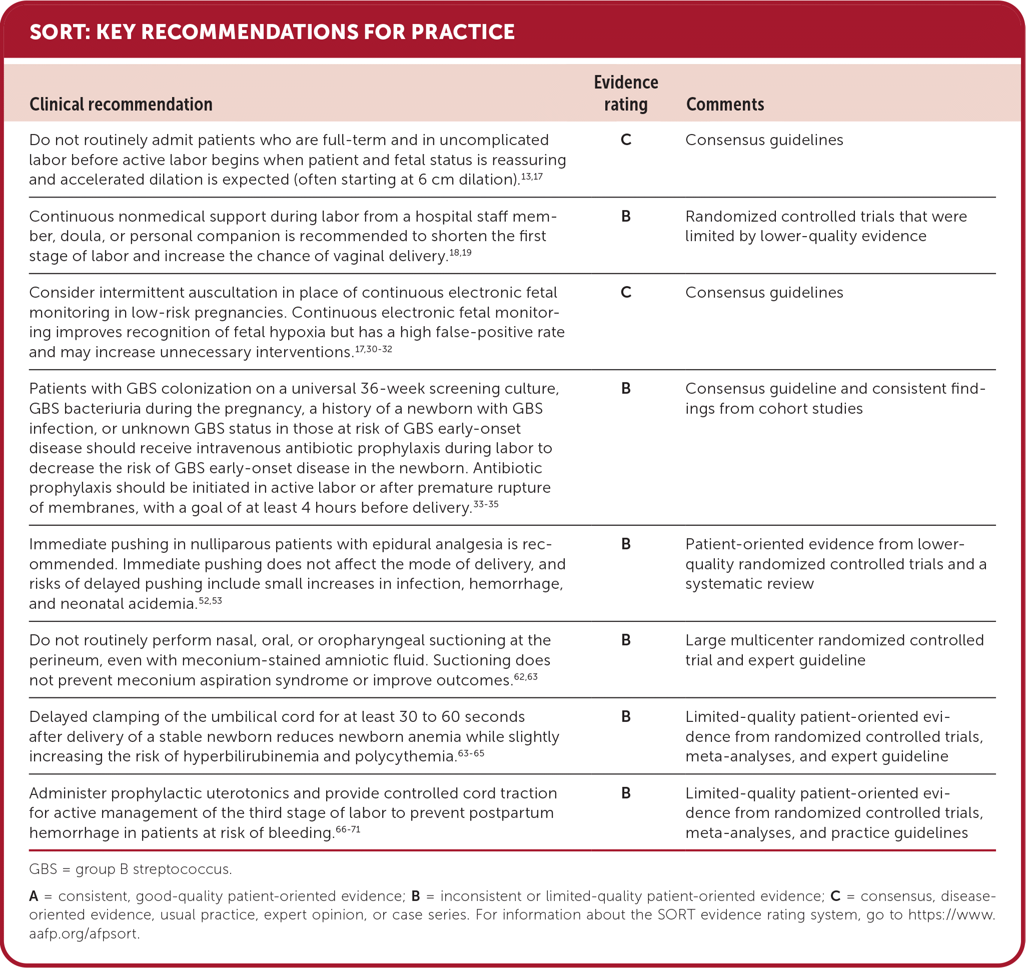
SORT: KEY RECOMMENDATIONS FOR PRACTICE
GBS = group B streptococcus.
A = consistent, good-quality patient-oriented evidence; B = inconsistent or limited-quality patient-oriented evidence; C = consensus, disease-oriented evidence, usual practice, expert opinion, or case series. For information about the SORT evidence rating system, go to https://www.aafp.org/afpsort.
BEST PRACTICES IN OBSTETRICS
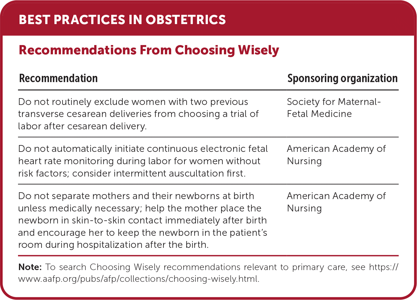
| Recommendation | Sponsoring organization |
|---|---|
| Do not routinely exclude women with two previous transverse cesarean deliveries from choosing a trial of labor after cesarean delivery. | Society for Maternal- Fetal Medicine |
| Do not automatically initiate continuous electronic fetal heart rate monitoring during labor for women without risk factors; consider intermittent auscultation first. | American Academy of Nursing |
| Do not separate mothers and their newborns at birth unless medically necessary; help the mother place the newborn in skin-to-skin contact immediately after birth and encourage her to keep the newborn in the patient's room during hospitalization after the birth. | American Academy of Nursing |
Note: To search Choosing Wisely recommendations relevant to primary care, see https://www.aafp.org/pubs/afp/collections/choosing-wisely.html.
TABLE 1. Guidelines for Redating Based on Ultrasonography
| Gestational age range* | Method of measurement | Discrepancy between ultrasound dating and last menstrual period dating that supports redating |
|---|---|---|
| ≤ 13 6/7 weeks ≤ 8 6/7 weeks 9 0/7 to 13 6/7 weeks | Crown-rump length | More than 5 days More than 7 days |
| 14 0/7 to 15 6/7 weeks | Biparietal diameter, head circumference, abdominal circumference, femur length | More than 7 days |
| 16 0/7 to 21 6/7 weeks | Biparietal diameter, head circumference, abdominal circumference, femur length | More than 10 days |
| 22 0/7 to 27 6/7 weeks | Biparietal diameter, head circumference, abdominal circumference, femur length | More than 14 days |
| 28 0/7 and greater weeks† | Biparietal diameter, head circumference, abdominal circumference, femur length | More than 21 days |
*—Based on last menstrual period.
†—Because of the risk of redating a small fetus that may be growth restricted, management decisions based on third-trimester ultrasonography alone are especially problematic and need to be guided by careful consideration of the entire clinical picture and close surveillance.
Reprinted with permission from Committee Opinion No. 700: methods for estimating the due date. Obstet Gynecol. 2017;129(5):e152.
The birthing process has the potential for psychological and physical morbidity. Patient autonomy must be respected, and patients should provide consent for each examination. Not every pregnant patient identifies as a female; attention should be given to respecting the patient's gender identity and pronouns.
There are only a few absolute contraindications to vaginal delivery, including an active genital herpes simplex outbreak, unknown or elevated HIV viral load, malpresentation, complete placenta previa, and a history of specific uterine incisions.4–6 Relative contraindications to vaginal delivery may be identified before labor, such as suspected fetal macrosomia or malpresentation of the first fetus in multiple gestation, or may present during labor, including labor dystocia or abnormal fetal heart tracing (Table 2).4–6
TABLE 2. Contraindications to Vaginal Delivery
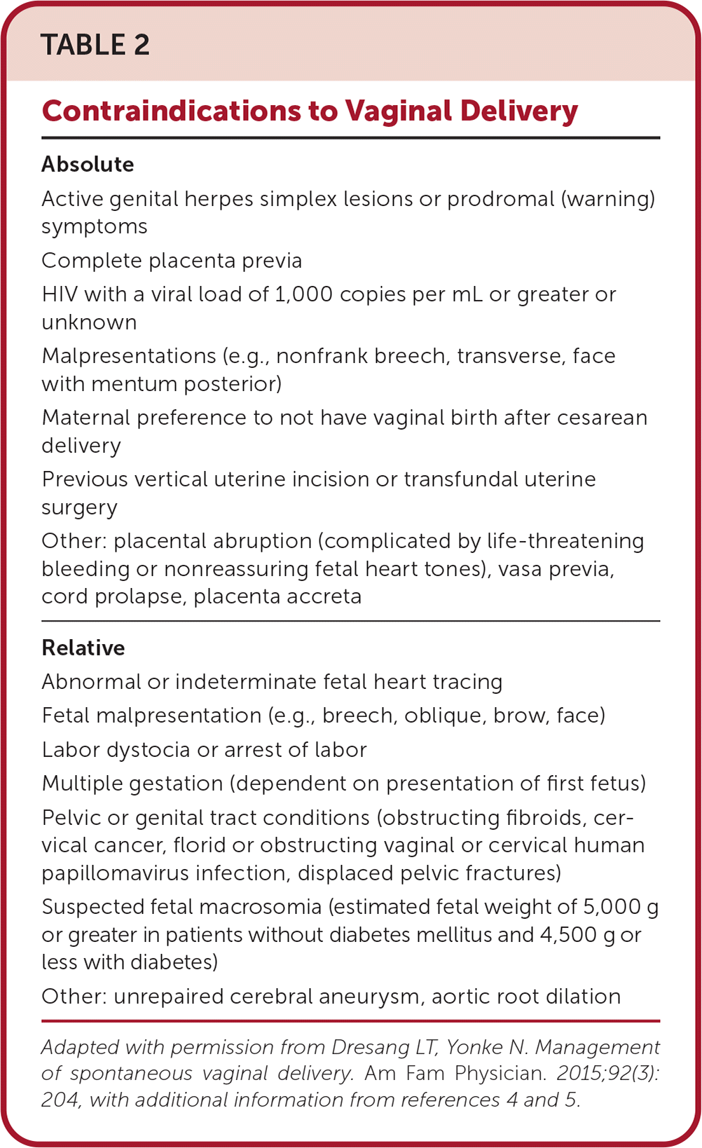
| Absolute |
| Active genital herpes simplex lesions or prodromal (warning) symptoms |
| Complete placenta previa |
| HIV with a viral load of 1,000 copies per mL or greater or unknown |
| Malpresentations (e.g., nonfrank breech, transverse, face with mentum posterior) |
| Maternal preference to not have vaginal birth after cesarean delivery |
| Previous vertical uterine incision or transfundal uterine surgery |
| Other: placental abruption (complicated by life-threatening bleeding or nonreassuring fetal heart tones), vasa previa, cord prolapse, placenta accreta |
| Relative |
| Abnormal or indeterminate fetal heart tracing |
| Fetal malpresentation (e.g., breech, oblique, brow, face) |
| Labor dystocia or arrest of labor |
| Multiple gestation (dependent on presentation of first fetus) |
| Pelvic or genital tract conditions (obstructing fibroids, cervical cancer, florid or obstructing vaginal or cervical human papillomavirus infection, displaced pelvic fractures) |
| Suspected fetal macrosomia (estimated fetal weight of 5,000 g or greater in patients without diabetes mellitus and 4,500 g or less with diabetes) |
| Other: unrepaired cerebral aneurysm, aortic root dilation |
Adapted with permission from Dresang LT, Yonke N. Management of spontaneous vaginal delivery. Am Fam Physician. 2015;92(3):204, with additional information from references 4 and 5.
Trial of Labor After Cesarean Delivery
Patients with a history of up to two transverse cesarean deliveries may be candidates for a trial of labor, depending on patient preference and patient and neonatal risk.7,8 The risk-based vaginal birth after cesarean calculator from the Eunice Kennedy Shriver National Institute of Child Health and Human Development can guide a shared decision-making discussion for candidates, with a minimum of 60% to 70% predicted success (https://www.mdcalc.com/calc/10433/vaginal-birth-after-cesarean-vbac).8,9 The discussion should include the increased risk of uterine rupture or dehiscence associated with a trial of labor after cesarean delivery and increased patient and neonatal morbidity if an urgent cesarean delivery becomes necessary.8 Because complications may be unpredictable, a trial of labor after cesarean delivery should only be pursued where resources for an emergency cesarean delivery are immediately available.8
Out-of-Hospital Birth
Patients may choose to have a vaginal delivery at home instead of in the hospital setting, often with the goal of reducing interventions.10 Although hospital births are safer, a patient's choice should be approached without judgment and with respect for autonomy over an important life event. A planned home birth is associated with an average absolute increased risk of perinatal death of up to 2 deaths per 1,000 patients.10 Home births are safest for appropriate low-risk candidates when assisted by licensed clinicians who meet standards set by the International Confederation of Midwives or who are integrated into a regulated health care system. Standardized guidelines for safe and timely transport to a nearby hospital should be used if needed.11
Stages of Labor
Spontaneous labor is when regular uterine contractions result in a cervical change via dilation or effacement and is divided into three stages (Table 3).12,13 If amniotic membranes rupture at term but before the onset of labor (i.e., premature rupture of membranes [PROM]), immediate labor induction reduces the time to birth and decreases infection risk without increasing cesarean delivery risk.14 For patients with reassuring fetal status after PROM, a limited period of expectant management (monitoring for labor progression without intervention) is also reasonable.15 Most patients begin spontaneous labor within 12 hours, and nearly all within 24 to 28 hours.15
TABLE 3. Stages of Labor
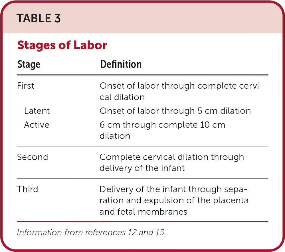
| Stage | Definition |
|---|---|
| First | Onset of labor through complete cervical dilation |
| Latent | Onset of labor through 5 cm dilation |
| Active | 6 cm through complete 10 cm dilation |
| Second | Complete cervical dilation through delivery of the infant |
| Third | Delivery of the infant through separation and expulsion of the placenta and fetal membranes |
FIRST STAGE
The first stage of labor progresses to complete cervical dilation and effacement.12 For most patients, the transition from the passive to active phase begins at 6 cm dilation.16 Active labor is consistently marked by accelerated cervical dilation, which is more variable at 6 cm or less.13,17 Patients at 37 weeks' gestation or later without maternal or neonatal concerns are not typically admitted to labor and delivery until reaching active labor.13,17 Admission during latent labor in the setting of maternal fatigue or a need for pain management may be appropriate.17
Interventions that may shorten the first stage of labor and increase the chance of vaginal delivery include continuous nonmedical support from a hospital staff member, doula, or personal companion; mobility, including walking, sitting, standing, and kneeling; and the use of a peanut ball (a peanut-shaped plastic ball placed between the knees of a person laboring in the lateral recumbent position) after epidural anesthesia.18–22 Amniotomy also shortens the duration of labor without affecting the mode of delivery or maternal infection risk compared with waiting for the spontaneous rupture of membranes.23
Although food intake has traditionally been restricted during labor to minimize gastric aspiration risk in case of emergent general anesthesia, this practice should be readdressed because the safety of anesthesia has advanced. Even though gastric emptying is delayed in pregnant patients, epidural analgesia appears to facilitate gastric emptying.24 A large review of obstetric analgesia complications did not find cases of aspiration during cesarean delivery, and a systematic review found that less restrictive food intake was associated with a slightly shorter duration of labor but similar operative rates without increased vomiting or other obstetric or neonatal harms.25,26
Assessment and Augmentation. Serial cervical examinations are performed throughout labor to assess cervical dilation, effacement, and fetal station.27 Frequent examinations may decrease the risk of cesarean delivery and neonatal morbidity27; however, cervical examinations are invasive and may cause discomfort. Examinations should be deferred until a change in management may be indicated, and unnecessary examinations should be avoided to reduce the risk of chorioamnionitis.28 Augmentation with pharmacologic stimulation of uterine contractions or artificial rupture of membranes to increase the frequency and strength of contractions may be indicated when the first stage of labor extends beyond the expected timeframe.29
Electronic Fetal Monitoring. Electronic fetal monitoring records the fetal heart rate in relation to maternal uterine contractions as a measure of fetal well-being during labor. Continuous electronic fetal monitoring is recommended in patients with prenatal or intrapartum risk factors for neonatal compromise.17,30,31 Although continuous electronic fetal monitoring improves the recognition of fetal hypoxia, use is limited by a high false-positive rate, which may increase unnecessary interventions, including instrumental vaginal birth and cesarean delivery. Continuous electronic fetal monitoring should not be used as the only diagnostic tool for intervention.30,32
The use of continuous electronic fetal monitoring is associated with a lower risk of neonatal seizures without affecting cerebral palsy risk or perinatal mortality.32 Patients at low risk of complications who want and consent to less frequent monitoring may be offered intermittent auscultation. This option for fetal heart rate assessment, which includes the use of a handheld Doppler every 5 to 15 minutes during labor, may depend on staff training and availability.17,31
Group B Streptococcus Prophylaxis. Group B streptococcus (GBS) is the most common cause of neonatal infection, with vertical transmission occurring in 50% of patients with colonization in the genitourinary or gastrointestinal tract.33 Universal screening for GBS colonization at 36 weeks' gestation via vaginal-rectal culture is recommended. Patients with a positive result on screening culture, GBS bacteriuria during the pregnancy, or a history of a newborn with GBS infection, even with a negative culture result during the current pregnancy, should receive intravenous antibiotic prophylaxis during labor to reduce the risk of early-onset GBS disease in the newborn.33 Prophylaxis is also indicated in patients with unknown GBS status and if risk factors for early-onset GBS disease are present (Table 4).33
TABLE 4. Indications for Intrapartum GBS Prophylaxis With Unknown GBS Status
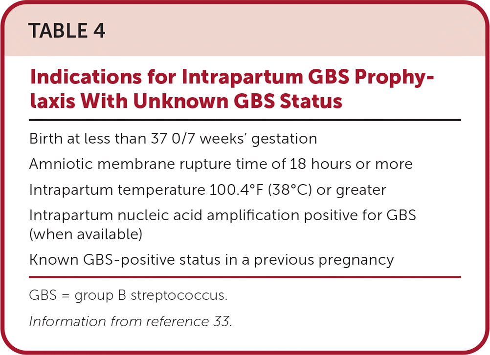
| Birth at less than 37 0/7 weeks' gestation |
| Amniotic membrane rupture time of 18 hours or more |
| Intrapartum temperature 100.4°F (38°C) or greater |
| Intrapartum nucleic acid amplification positive for GBS (when available) |
| Known GBS-positive status in a previous pregnancy |
GBS = group B streptococcus.
Information from reference 33.
Antibiotic prophylaxis is initiated in active labor or following PROM and continued through delivery. It is most effective when administered for at least 4 hours before delivery, although 2 hours of administration reduces vaginal colony counts and the risk of neonatal sepsis.33–35 Intravenous penicillin is the preferred antibiotic for prophylaxis; second-line antibiotics may be required for patients with a severe immunoglobulin E–mediated penicillin allergy based on GBS susceptibility testing.33
Pain Management. Labor is a painful process. Because empowerment improves patient satisfaction, and analgesia does not affect the mode of delivery, pain should be assessed frequently and managed based on patient preference.36–38 Nonpharmacologic options such as water immersion, pelvic motion exercises, lumbosacral massage, and sterile water injections can help ease pain and delay or reduce the use of pharmacologic analgesia.39–42
Nonopioid medications for pain management, including antihistamines, sedatives, and acetaminophen, are more satisfactory than placebo but provide less pain relief than opioids.43 Intramuscular or intravenous opioids provide moderate pain relief but can be associated with adverse effects, including maternal nausea, vomiting, and drowsiness.44 Opioids cross the placenta and can cause newborn respiratory depression when administered closer to the time of delivery.38 Neuraxial analgesia, via epidural or spinal techniques, results in better pain control than systemic opioids and is associated with fewer adverse effects.38,45 Self-administered inhaled nitrous oxide reduces pain intensity without limiting mobility, with similar maternal adverse effects as systemic opioids.46 A previous article in American Family Physician provides more information about analgesia during labor.42
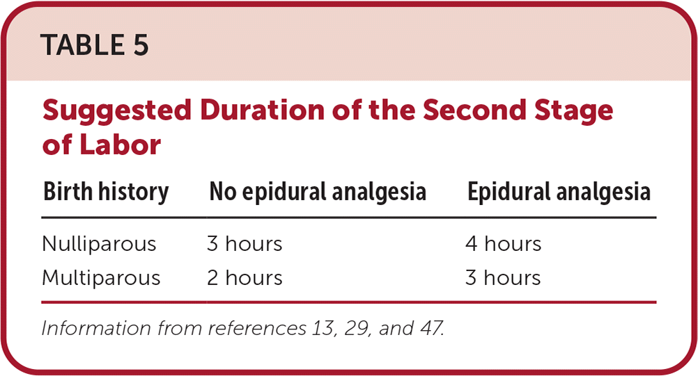
SECOND STAGE
The second stage of labor proceeds from complete cervical dilation to delivery of the infant.12 During this stage, the fetus progresses through head engagement with the maternal pelvis, head flexion, descent through the birth canal, internal rotation, head extension, external rotation or restitution, and expulsion. Although the duration of the second stage is variable, suggested maximum durations based on parity and analgesia are provided in Table 5.13,29,47 Longer durations may be appropriate with documented fetal descent; however, the chance of successful spontaneous vaginal delivery decreases with each additional hour.5,16
TABLE 5. Suggested Duration of the Second Stage of Labor
Fetal Head Position. According to a small cohort study, the occiput posterior is the most common fetal head position within the maternal pelvis during the first stage of labor. Most fetuses have rotated to the occiput anterior position by complete cervical dilation.48 Manual rotation of the fetal head in persistent occiput posterior position during the second stage modestly increases the rate of spontaneous vaginal delivery and decreases the use of episiotomy 49; the success of this maneuver improves when the fetal spine location is identified by ultrasonography.50 Maternal hands and knees position during labor may improve back pain but does not result in rotation of the fetal head.51
Pushing Strategies. Historically, delaying pushing by 1 or more hours after complete dilation was thought to decrease active pushing time and increase the rate of spontaneous vaginal delivery in nulliparous patients with epidural analgesia. A newer systematic review demonstrated that delayed pushing does not increase vaginal delivery rates compared with immediate pushing.52 Risks of delayed pushing, including a small increase in infection, hemorrhage, and neonatal acidemia, should be shared with patients before initiating this approach.52,53
There is little evidence to guide positioning and pushing. In a large trial of nulliparous patients with epidural analgesia, patients who chose to push lying on their side were more likely to deliver vaginally than those in upright positions.54 For patients without epidural analgesia, vertical positioning may shorten the second stage of labor at the risk of higher blood loss.55 There is no difference between coached and spontaneous pushing on the duration or mode of delivery.56,57 Patients without other clinical concerns should be encouraged to change positions and use their preferred pushing technique.
Delivery Technique. Applying warm packs to the perineum during the second stage of labor reduces perineal lacerations, including second- and third-degree lacerations, episiotomy rates, and postpartum pain.58 Selective use of episiotomy in an unassisted vaginal birth reduces perineal trauma compared with routine episiotomy.59
Delivery of the fetus is imminent at the time of +5 fetal station (crowning). The physician's dominant hand should support the fetal head and maintain its flexed position, while the other hand supports the perineum by squeezing the lateral perineal tissue toward the midline. Encouraging small pushes allows controlled delivery of the fetal head.
After delivery of the jaw, the fetal neck should be examined for the presence of a nuchal cord. If not overly tight, a nuchal cord may be reduced at the perineum or after delivery. Following delivery of the head, gentle downward or axial traction allows for the delivery of the anterior shoulder under the maternal pubic symphysis. Gentle upward traction allows for delivery of the posterior shoulder and the rest of the fetus. Shoulder dystocia (i.e., failure of the anterior shoulder to deliver with gentle axial traction) is an obstetric emergency and requires immediate intervention.60
Initial Newborn Management. For infants who do not need resuscitation, delivery directly onto the mother's chest allows for the immediate initiation of skin-to-skin care, which increases bonding, improves neonatal transition, and improves breastfeeding outcomes.61 Routine nasal, oral, or oropharyngeal suctioning is not recommended after delivery.62,63 In the setting of meconium-stained amniotic fluid, oropharyngeal suctioning does not prevent meconium aspiration syndrome or improve outcomes, but a credentialed and skilled neonatal resuscitation team should be available.63
Delayed clamping of the umbilical cord for at least 30 to 60 seconds after delivery of a stable infant reduces newborn anemia while slightly increasing the risk of hyperbilirubinemia and polycythemia.63–65 Following this delay, the umbilical cord is cut with sterile scissors between two clamps to separate the infant from the placenta.
THIRD STAGE
The third stage of labor proceeds from delivery of the infant through delivery of the placenta.12 This stage generally lasts 30 minutes or less.
Reducing Postpartum Hemorrhage. Postpartum hemorrhage is the most common cause of maternal morbidity and mortality worldwide.66 Postpartum hemorrhage is defined as blood loss greater than 1,000 mL or less than 1,000 mL with signs or symptoms of hypovolemia in the first 24 hours following delivery. Active management of the third stage is the standard of care to reduce postpartum hemorrhage and lower the risk of maternal hemoglobin levels of less than 9 g per dL (90 g per L) postpartum. However, less evidence supports this approach in patients with a low risk of bleeding.67
Active management includes prophylactic uterotonic administration and controlled traction on the umbilical cord.66,67 Intravenous or intramuscular oxytocin is an effective and well-tolerated uterotonic medication.66,68 Oxytocin combined with methylergonovine or misoprostol is more effective than oxytocin alone in preventing blood loss greater than 500 mL, with increased adverse effects.69 Although uterotonic administration before delivery of the placenta may reduce postpartum hemorrhage while increasing retained placenta, evidence is insufficient.70 Some sources also recommend early cord clamping and uterine massage as active third-stage management components.66,67 Early cord clamping may reduce postpartum hemorrhage; however, evidence is insufficient.67 Similarly, the importance of uterine massage is unproven.66
Delivery of the Placenta. Controlled cord traction, with one hand applying traction on the cord and the other providing suprapubic pressure on the uterus toward the umbilicus, reduces manual placental removal without affecting postpartum hemorrhage.71
Although based on very low-certainty evidence, umbilical vein injection with oxytocin may decrease the need for manual placenta removal when placental delivery is delayed.72 If the placenta has not been delivered within 30 minutes, manual removal of the placenta with a gloved hand may be required. The placenta should be inspected to ensure that it has been removed intact since a retained placenta can lead to postpartum hemorrhage and infection. There is no evidence to support administration of prophylactic antibiotics to prevent endometritis following the manual removal of the placenta.73
Perineal Lacerations
Before and after placental delivery, the vagina and perineum should be inspected for lacerations, including obstetric anal sphincter injuries. Lacerations that alter anatomy or are not hemostatic should be repaired. Techniques for laceration repair are discussed in a previous American Family Physician article.74
This article updates previous articles on this topic by Dresang and Yonke6 and Patterson, et al.75
Data Sources: A search was completed in Essential Evidence Plus, POEMs, the Cochrane database, and the National Institute for Health and Care Excellence guidelines using the key terms spontaneous vaginal delivery with systematic reviews, practice guidelines, diagnosis, and therapy. Search dates: April to July 2023; October to December 2023; and March 2024.
