

A 40-year-old woman presented with a painful lesion on her thigh. It had formed 3 months earlier as a red spot and gradually darkened. She had no other lesions.
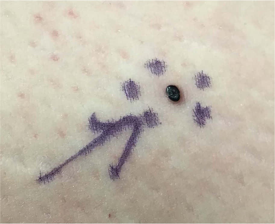
The examination revealed a black papule on her inner thigh with a diameter of 2 mm (Figure 1). Dermoscopic examination showed dark purple lacunes. No other similar lesions were found during the full-body skin examination, including the oral cavity and tongue. An urgent excision of the lesion was performed.
FIGURE 1
QUESTION
Based on the patient's history and physical examination, which one of the following is the most likely diagnosis?
- A. Pyogenic granuloma.
- B. Solitary angiokeratoma.
- C. Superficial spreading malignant melanoma.
- D. Verrucous hemangioma.
DISCUSSION
The answer is B: solitary angiokeratoma. This rare benign vascular lesion can occur anywhere on the body but is most common on the lower extremities. Men are most likely to develop these lesions, and prevalence increases with age. The incidence of solitary angiokeratoma is difficult to determine because the lesions are often asymptomatic.1 Multiple angiokeratomas involving the scrotum or vulva are called angiokeratomas of Fordyce. Multiple acral verrucous angiokeratomas in children are known as angiokeratomas of Mibelli. Angiokeratoma circumscriptum presents at birth as a cluster of angiokeratomas involving the lower extremities.2
Angiokeratomas are sporadic, with no hereditary origin or significant predisposing factors. Eruptive angiokeratomas have been rarely reported in patients taking enoxaparin.3 Fabry disease, a rare X-linked genetic disorder, features multiple angiokeratomas in a bathing suit distribution (on the thighs and buttocks) and is associated with renal, heart, and neurologic symptoms.4
Diagnosis of angiokeratoma is clinical and can be aided by dermoscopy. Dermoscopic features include dark lacunae, a whitish veil, keratinization, and hemorrhagic crusts. Most solitary angiokeratomas do not require treatment. However, treatment can be offered when a patient has a significant cosmetic burden, bleeding, or thrombosis causing inflammation and pain. Treatment options depend on the size and location of the lesion and patient preference. Surgical excision is usually the most effective approach and has a low recurrence rate. Other treatment options include cautery, laser therapy, or cryotherapy.5
Pyogenic granuloma is a benign, rapidly growing vascular tumor that presents as a fleshy, red papule or nodule. The tumor tends to bleed easily with minor trauma and commonly occurs on the fingers, gingiva, and lips.6
Superficial spreading malignant melanoma is the most prevalent form of malignant melanoma, accounting for 60% to 70% of cases. It is characterized by irregular pigmented macules or patches that typically develop in areas exposed to intense intermittent sunlight. It is most common on the legs in females and the trunk in males. Superficial spreading of malignant melanoma is suggested by slowly growing or changing flat patches or macules with any of the ABCDE characteristics: asymmetrical shape or color, border irregularity, color variation and change, the presence of a different mole (ugly duckling sign), and a history of evolution.7
Verrucous hemangioma is a vascular lesion characterized by verrucous and keratotic plaques or nodules. It is typically present at birth or develops during early childhood and primarily involves the lower extremities, making it important to consider in the differential diagnosis of vascular lesions in this area.8
Read the full article
Get immediate access, anytime, anywhere.
Choose a single article, issue, or full-access subscription.
Earn up to 14 CME credits per issue.
