

A 50-year-old man with end-stage renal disease presented with an intensely pruritic rash on his upper and lower extremities, abdomen, and back. The rash had appeared spontaneously three months before the visit and was not associated with systemic symptoms. The patient had a history of type 2 diabetes mellitus and was receiving dialysis.
Physical examination revealed numerous papules of various sizes (Figure 1). Each lesion had a central depression and an erythematous halo. The rash featured hyperkeratosis but no fluctuance or induration. A punch biopsy of a lesion on the upper right quadrant of the abdomen was performed. It revealed a keratin-filled channel in the shape of a funnel with histocyte-rich infiltrate and fibrosis at the base of the invagination. Grocott-Gomori methenamine–silver nitrate and periodic acid–Schiff stains were negative for fungal elements.
FIGURE 1
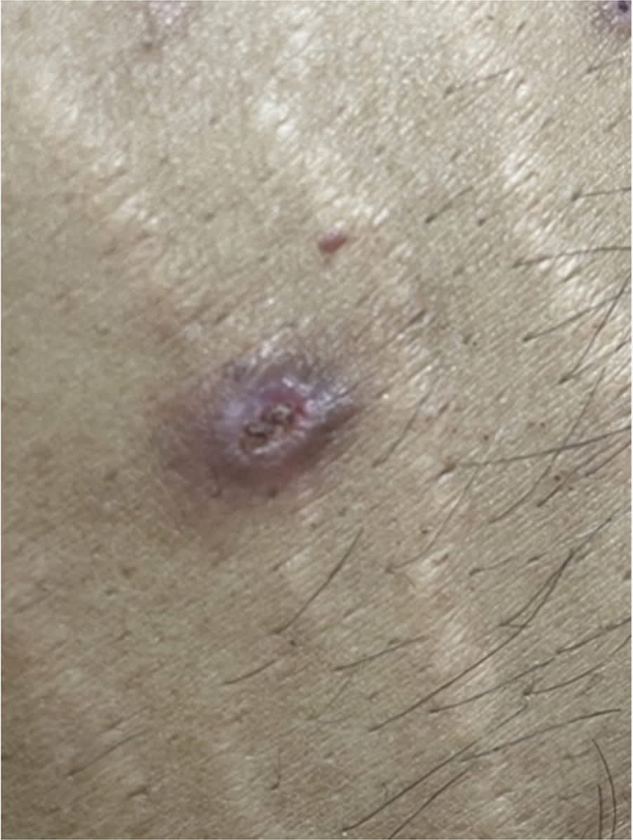
QUESTION
Based on the patient's history and physical examination, which one of the following is the most likely diagnosis?
- A. Acquired perforating dermatosis.
- B. Dermatofibroma.
- C. Keratosis follicularis.
- D. Molluscum contagiosum.
- E. Prurigo nodularis.
DISCUSSION
The correct answer is A: acquired perforating dermatosis, a skin condition caused by transepidermal elimination of connective tissue.1 The disorder is associated with chronic systemic diseases, most commonly diabetes and chronic renal failure requiring dialysis.2 Diagnosis is made by clinical and histopathologic findings. The diagnosis is challenging because clear identification of connective tissue deposits can be difficult.2
The rash most commonly erupts on the lower extremities, but it can also be widespread. The lesions are typically round and umbilicated, with nodules or a central keratotic core. They may be erythematous, hyperpigmented, or the same color as the surrounding skin.1 Due to extreme pruritus, the keratotic core is often removed by scratching.2 The clinical course is linked to treatment of the underlying disease. Treatment for acquired perforating dermatosis is challenging because no clinical trials of treatments have been performed. Most treatment options are based on case reports, and they typically use retinoids, topical and intradermal corticosteroids, and ultraviolet B phototherapy.1
Dermatofibromas, also known as cutaneous fibrous histiocytomas, are benign tumors of dermal origin.1 The diagnosis is made clinically. The patient may recall trauma to the area, such as an insect bite or shaving injury. On physical examination, the nodules are typically solitary, oval, painless, firm, smooth, and of various sizes. Coloration varies; dermatofibromas can be violaceous, brown, or the same color as the surrounding skin. They exhibit a dimple sign or Fitzpatrick sign (i.e., dimpling on lateral compression).3
Keratosis follicularis, also known as Darier disease, is an inherited autosomal dominant condition.4 Clinical manifestations usually appear between 6 and 20 years of age but occasionally occur in older adults. Typically, the first clinical manifestations of the disease are on the hands and nails. Lesions are usually crusted, pink papules that coalesce into plaques. As the condition progresses, lesions develop in intertriginous areas and on the scalp. The papules have a foul-smelling odor. Symptoms are relapsing, remitting, and exacerbated by heat.
Molluscum contagiosum most commonly occurs in children, although it is also possible in immunocompromised adults. The lesions are caused by poxvirus and are spread via direct physical contact. The papules are 1 to 3 mm, round, often pink and pearly, and have a central area of umbilication.5
Read the full article
Get immediate access, anytime, anywhere.
Choose a single article, issue, or full-access subscription.
Earn up to 14 CME credits per issue.
