

A 20-year-old man presented with right shoulder pain. He had been shoveling snow when his shovel struck a patch of ice, and he immediately felt pain in his shoulder. When he attempted to raise his arm, he noticed pain and clicking sensations. His medical history was significant for right clavicle fractures 12 and 4 years previously. Both injuries were managed nonoperatively, and normal function was restored.
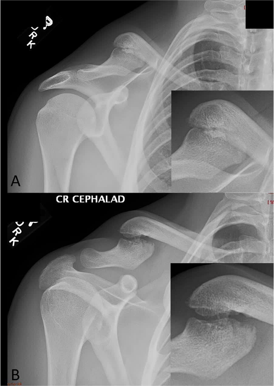
Physical examination revealed a notable deformity of the right clavicle (Figure 1). The patient had normal right upper extremity strength with normal distal sensation and pulses. The deformity had minimal tenderness, but the patient reported pain with the passive cross-arm test (passive horizontal shoulder adduction). Plain radiography was performed (Figure 2).
FIGURE 1
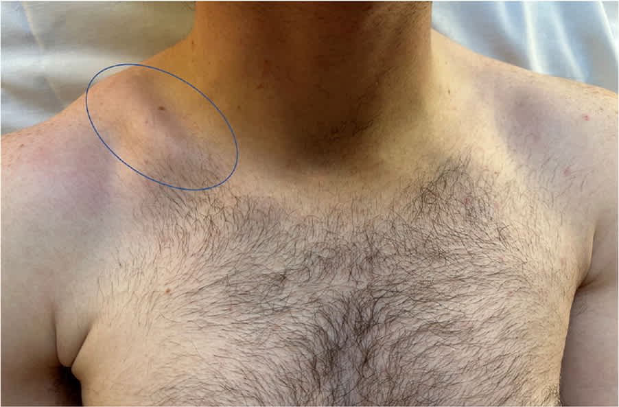
FIGURE 2
QUESTION
Based on the patient's history, physical examination, and plain radiography, which one of the following is the most likely diagnosis?
- A. Acute nondisplaced fracture.
- B. Nonunion fracture.
- C. Pathologic fracture.
- D. Stress fracture.
DISCUSSION
The answer is B: nonunion fracture, which occurs when a previous fracture does not heal within 4 to 6 months. It develops in 10% to 20% of displaced midshaft clavicle fractures.1 Clavicle fractures represent about 2.6% of all fractures and are usually caused by a fall directly onto the lateral shoulder.2–4
Differentiating an acute fracture from a nonunion fracture in patients with a prior clavicle fracture can be difficult.1,2 This patient had a long-standing nontender clavicle deformity. Smooth, well-rounded, and well-corticated fracture margins on radiographs indicate chronicity and confirm the diagnosis of nonunion fracture.1,2
Acute clavicle fractures are typically due to trauma and feature pain, tenderness, and deformity on examination. Most midshaft fractures are displaced and comminuted.2–5 Radiographic features of an acute nondisplaced fracture include cortical disruption with sharp edges.2 Risks of surgical management include infection, surgical scarring, hardware impingement, and the need for secondary procedures.2,5 Compared with operative management, conservative management is associated with visible clavicle deformity and higher nonunion fracture rate.2,5
Atraumatic or minimally traumatic fractures include pathologic and stress fractures, which are caused by focal bone weakness or repetitive stress. Pathologic fractures occur due to a local or systemic process weakening or altering the bone structure.2,6 The process can be benign or malignant; cystic lesions are the most common underlying etiology.2 Physical examination findings are similar to those of an acute traumatic fracture with tenderness at the fracture. Radiographic patterns reveal the underlying abnormal structure with an acute fracture pattern.2,6
Stress fractures are overuse injuries caused by repetitive microtrauma to a long bone and rarely occur in the clavicle.7,8 On physical examination, the area is tender, with no obvious deformity. Plain radiography findings usually remain negative for weeks but may show faint cortical radiolucency.2,6,7 A periosteal reaction with cortical thickening typically appears later. Signs of new bone formation, such as linear sclerosis, may appear before a cortical break with a lucent fracture line. Advanced imaging may be necessary to confirm the diagnosis.2,6,8
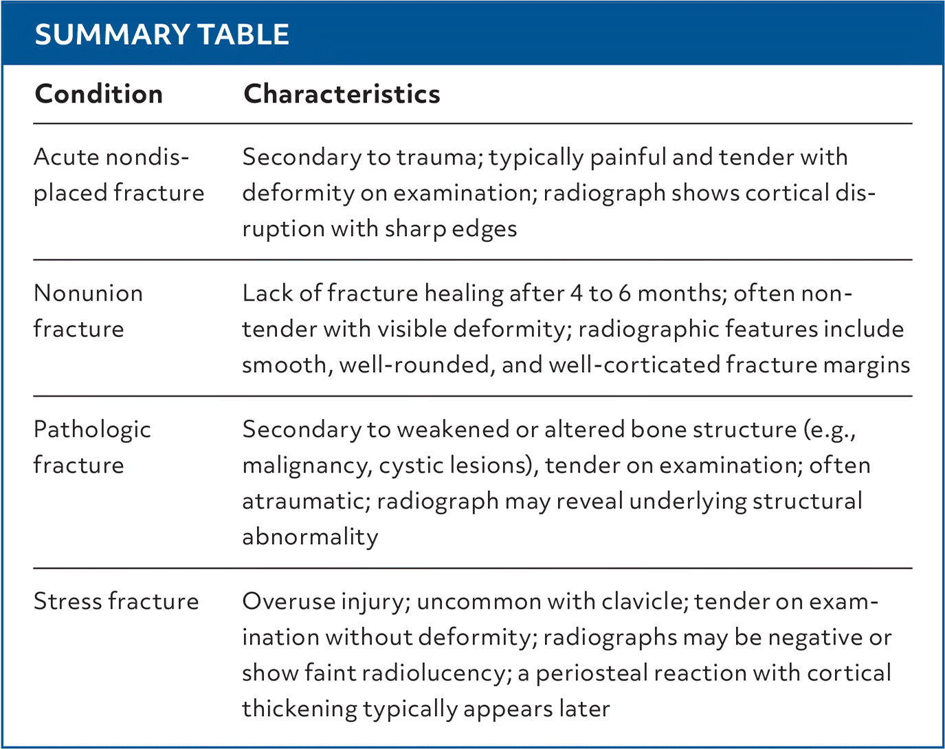
SUMMARY TABLE
| Condition | Characteristics |
|---|---|
| Acute nondisplaced fracture | Secondary to trauma; typically painful and tender with deformity on examination; radiograph shows cortical disruption with sharp edges |
| Nonunion fracture | Lack of fracture healing after 4 to 6 months; often non-tender with visible deformity; radiographic features include smooth, well-rounded, and well-corticated fracture margins |
| Pathologic fracture | Secondary to weakened or altered bone structure (e.g., malignancy, cystic lesions), tender on examination; often atraumatic; radiograph may reveal underlying structural abnormality |
| Stress fracture | Overuse injury; uncommon with clavicle; tender on examination without deformity; radiographs may be negative or show faint radiolucency; a periosteal reaction with cortical thickening typically appears later |
Read the full article
Get immediate access, anytime, anywhere.
Choose a single article, issue, or full-access subscription.
Earn up to 14 CME credits per issue.
